
Diseases of the sinus node (SAN) are common leading to bradycardia requiring electronic pacemaker implantation.1 To avoid problems associated with such devices efforts have been made to develop biological pacemakers by ion channel expression in the myocardium.2 Our aim is to apply this concept to the diseased SAN. Wistar rats were killed humanely, the SAN was dissected as previously described.3 Control (n=4) and ‘sick sinus’ preparations (inferior portion after division below level of leading pacemaker site; n=4) were then cultured. Electrical activity was monitored using 0.15 mm stainless steel electrodes. Tissue was frozen and sections prepared, Masson’s trichrome and immunostaining (caveolin 3) were performed as previously described.4 The sick sinus model displayed a slower rate than the control, and both slowed over a five day period (170 ± 12.5 to 124 ± 10.8 beats per minute (bpm) and 277 ± 9.7 to 211 ± 13.4 bpm respectively [mean ± SEM]). No histological changes were seen with Masson’s trichrome staining (n=3) and there was no change in cell size as assessed with Caveolin3 immunostaining (n=10). Further preparations (n=2) were injected with 1 x 10 8 infectious units of adenovirus Ad5-PREP-lacZ and expression of β-galactosidase was assessed by X-gal staining after 48 h. Tissue sections were prepared and immunostaining (ANP and HCN4) performed as above. X-gal staining of whole SAN infected with Ad5-PREP-lacZ showed transgene expression at injection sites in the superior and mid SAN. 15 μm X-gal stained sections revealed high levels of lacZ expression in the SAN area. Immunostaining for ANP (negative marker for SAN), and HCN4 (positive marker for SAN) confirmed transgene expression was in the SAN (figure 1). This method provides a useful in vitro model of pathological bradycardia and furthermore the tissue is receptive to adenovirus infection with high levels of transgene expression in the SAN. This provides proof of concept for the use of this model to study sinus node biopacemaking as a potential treatment for sick sinus syndrome.
University of Manchester (2007) Proc Physiol Soc 8, PC36
Poster Communications: Adenovirus-mediated transgene expression in the sinus node and development of a bradycardic model for the in vitro study of biopacemaking in sick sinus syndrome
G. M. Morris1, M. R. Boyett1, P. A. Kingston2, M. Lei1, H. Dobrzynski1
1. Cardiac Electrophysiology, University of Manchester, Manchester, United Kingdom. 2. Vascular Gene Therapy Unit, University of Manchester, Manchester, United Kingdom.
View other abstracts by:
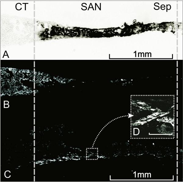
Figure 1. Adjacent sections prepared from rat SAN following injection with 1x10⁸ infectious units of Ad5-PREP-lacZ with tissue culture for 48 h. A, β-galactosidase expression assessed by X-gal staining (dark area) (CT, crista terminalis; Sep, interatrial septum). B, C, immunolabelling for ANP (B) and HCN4 (C). D, high power image of immunolabelling for HCN4 in the area indicated by the dotted box. The white areas in b, c and d represent positive labelling; scale bar in box d is 100 μm.
Where applicable, experiments conform with Society ethical requirements.