
High-resolution three-dimensional images of myocytes and collagen have been acquired across the left ventricular wall in ten normal Wistar-Kyoto (WKY) and thirteen spontaneously hypertensive (SHR) rat hearts using extended-volume confocal microscopy,1 with at least 2 hearts of each strain imaged at each age endpoint of 3, 12, 18 and 24 months. Tissue blocks were prepared as described previously2 and imaged at 1 µm resolution. The resulting 3-D image volumes were approximately 4 mm × 1 mm × 0.3 mm with isometric voxels of size (1 µm)3. A higher resolution image volume (560 µm × 560 µm × 330 µm, (0.4 μm)3 voxel size) was also acquired from the midwall of one heart of each strain at 12 months of age. Collagen volume fractions were computed on each block using a modified Top-hat filter that identifies regions that are bright relative to the local background. Specific myocardial structural components (myocytes, blood vessels, interlaminar spaces and different collagen structures (endomysial, perimysial, perivascular, scar)) were segmented in the midwall of each image volume by digitally tracing structural boundaries. Laminar width was measured along lines perpendicular to the myolaminae in the midwall region at 200 µm intervals. To identify laminar remodelling independent of changes in cell size, the laminar width was normalised by the average myocyte diameter in each block. Results are presented in Figure 1, where the collagen fractions (A) and layer widths (B) are expressed as group mean ± SEM. The myocyte areas (C) are expressed as box plots (first, median and third quartiles, with whiskers at 10% and 90%) to avoid making assumptions about the underlying statistical distribution. Subsequent analysis was performed using a 2-factor ANOVA with statistical significance based on a value of P ≤ 0.05. Collagen fraction increases with age from a similar baseline in both strains, but is more pronounced in the SHR. Myocyte cross-sectional area increases with age in the SHR, but not in the WKY. Both of these measures are significant for strain and strain-age interaction. The layer width in terms of myocyte diameters shows a significant increase at 18 and 24 months in the SHR (significant for strain, age, and strain-age interaction). It is clear that significant morphologic changes occur in the tissue structure of the SHR around 12 months with increasing collagen and remodelling of the laminar organisation. High-resolution analysis of the change in collagen at this time-point indicates that the increase is due to both scarring and increasing endomysial collagen. While some of this change occurs naturally with age, it is significantly accelerated in the SHR.
University College Dublin (2009) Proc Physiol Soc 15, C27
Oral Communications: Age-Related Changes in Left-Ventricular Structure in the Spontaneously Hypertensive Rat
G. B. Sands1, A. J. Pope2, B. H. Smaill1,2, I. J. LeGrice1,2
1. Bioengineering Institute, University of Auckland, Auckland, New Zealand. 2. Department of Physiology, University of Auckland, Auckland, New Zealand.
View other abstracts by:
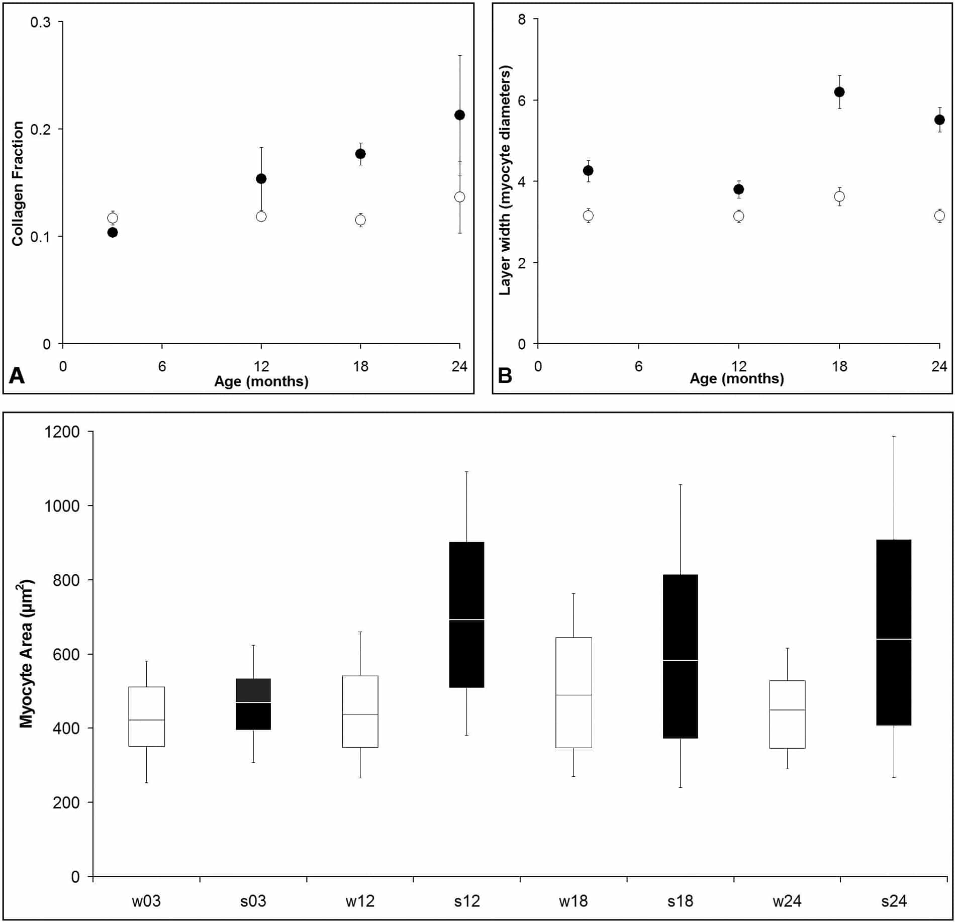
Figure 1. Comparison of left-ventricular tissue structure in SHR (dark, filled) and WKY (white, open) hearts. (A) Collagen fraction. (B) Layer width, measured as number of myocyte diameters. (C) Myocyte cross-sectional area.
Where applicable, experiments conform with Society ethical requirements.