
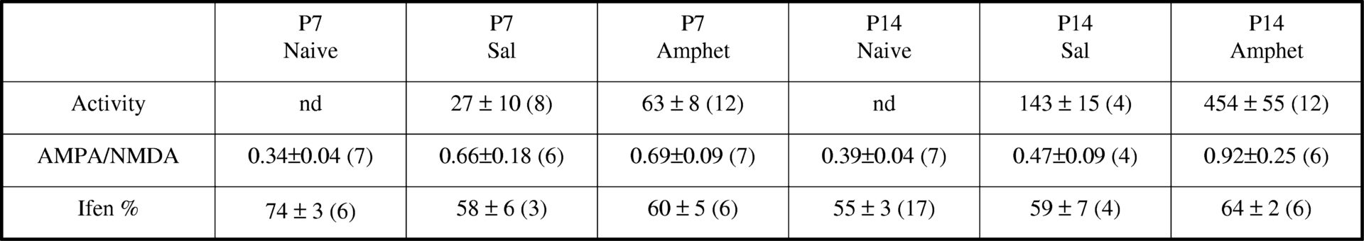
Glutamatergic synapses in rodent midbrain dopaminergic neurones show an increase in the ratio of AMPA receptor (AMPAR)-mediated excitatory postsynaptic currents (EPSCs) to NMDA receptor (NMDAR)-mediated EPSCs following a single dose of amphetamine administered 2-24 hours prior to EPSC recordings (Saal et al., 2003; Faleiro et al., 2004). This form of synaptic plasticity may contribute to behavioural adaptations seen in response to amphetamine, and may reflect changes in synaptic AMPAR expression, NMDAR expression, or both. Previously we have shown that synaptic NMDARs in rat substantia nigra dopaminergic neurones contain both NR2B and NR2D subunits (Brothwell et al., 2008). We have used whole-cell patch-clamp recordings to determine whether or not the sensitivity of NMDARs to the NR2B-preferring antagonist, ifenprodil, is changed in dopaminergic neurones following a single dose of amphetamine. Rats aged ~postnatal day (P)6 or ~P13 were given a single intra-peritoneal injection of amphetamine (2.5 mg kg-1) or saline control, and the locomotor response for 45-60 minutes following the injection was computed (number of infrared beam breaks); locomotor activity was significantly greater (P<0.05, t-test) in rats injected with amphetamine compared with saline at the two ages tested (Table 1; mean ± SEM, n in parentheses). Approximately 24 hours later, midbrain slices containing substantia nigra were prepared. In dopaminergic neurones voltage-clamped to +40mV (to remove Mg2+ block of NMDARs), EPSCs were evoked by electrical stimulation in the presence of 10 μM glycine. In the first set of experiments the total EPSC, consisting of AMPAR- and NMDAR-mediated components, was recorded and then D-AP5 (50 μM) applied to block NMDAR-EPSCs. The AMPAR-EPSC / NMDAR-EPSC ratio was significantly greater (P<0.05; ANOVA) in rats aged ~P14 injected with amphetamine compared with naïve rats (Table 1). In the second set of experiments NMDAR-EPSCs were pharmacologically isolated and the effect of ifenprodil (10 μM) was determined. There was no significant difference in the inhibition of NMDAR-EPSCs by ifenprodil in rats injected with amphetamine (Table 1). These results suggest that the change in the AMPAR-EPSC / NMDAR-EPSC ratio is developmentally sensitive, and that the proportion of synaptic NR2B-containing NMDARs is not altered by a single dose of amphetamine.
University College Dublin (2009) Proc Physiol Soc 15, PC12
Poster Communications: Amphetamine administration evokes no change in ifenprodil-sensitivity of synaptic NMDA receptors in dopaminergic neurones of rat substantia nigra
F. Suarez1, M. Smith1, A. J. Gibb2, S. Jones1
1. Department of Physiology, Development and Neuroscience, University of Cambridge, Cambridge, United Kingdom. 2. Department of Neuroscience, Physiology and Pharmacology, University College London, London, United Kingdom.
View other abstracts by:
Where applicable, experiments conform with Society ethical requirements.