
Basic and clinical studies have shown that bone marrow cell therapy can improve cardiac function following infarction (1). In experimental animals, reported stem cell-mediated changes range from no measurable improvement to the complete restoration of function (2), whereas in the clinic the average improvement in left ventricular ejection fraction is 2-3% (3). A possible explanation for the discrepancy between basic and clinical results is that few basic studies have used the MRI methods that were used in clinical trials for measuring cardiac function. Consequently, we employed cine MRI to determine the effect of bone marrow stromal cells (BMSCs) on cardiac function in rats. Wistar rat BMSCs were isolated from tibias and femurs, cultured for 21 days, characterized using flow cytometry and labeled with iron oxide particles and a fluorescent marker to allow in vivo cell tracking and ex vivo cell identification, respectively (4). Neither label affected in vitro cell proliferation or differentiation. Wistar rats were anaesthetised with 2.5% isoflurane in O2 and the coronary artery was occluded. BMSCs or control media were either injected into the infarct periphery 10 minutes after infarction (n = 34) or infused systemically 1 or 3 days after infarction (n = 30). MRI was used to measure cardiac morphology and function and to determine cell distribution for 10 weeks after infarction and cell therapy. In vivo MRI, histology and cell re-isolation confirmed successful BMSC delivery and retention within the myocardium throughout the experiment (Figure). However, no significant improvement in any measure of cardiac morphology or function was observed at any time (Table). We conclude that cultured BMSCs are not the optimal cell population to treat the infarcted heart.
University of Cambridge (2008) Proc Physiol Soc 11, C58
Oral Communications: Bone marrow-derived stromal cells home to and remain in the infarcted rat heart, but fail to improve function: an in vivo MRI study
D. J. Stuckey1, C. A. Carr1, L. Tatton2, D. J. Tyler1, S. J. Hale2, D. Sweeney2, J. E. Schniedar1, E. Martin-Rendon2, G. K. Radda1, S. E. Harding3, S. M. Watt2, K. Clarke1
1. Department of Physiology, Anatomy and Genetics, University of Oxford, Oxford, United Kingdom. 2. National Blood Service, Oxford, United Kingdom. 3. Imperial College, London, United Kingdom.
View other abstracts by:
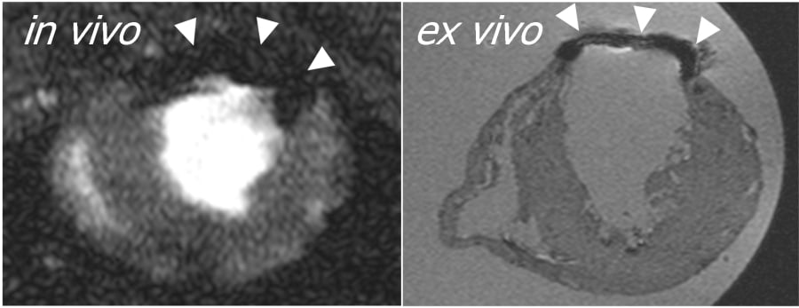
In vivo and ex vivo MR images of an infarcted rat heart. Arrows indicate position of BMSC engraftment
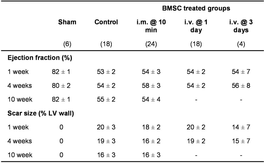
Ejection fractions and scar sizes measured using in vivo MRI up to 10 weeks after myocardial infarction and BMSC administration. Mean ± sem.
Where applicable, experiments conform with Society ethical requirements.