
Mild brain injuries (MBIs) in boxing are a major public concern due to the risk of accumulative effects. The severity of traumatic brain injury is associated with the early post-traumatic release of protein S-100B and neurone specific enolase (NSE). Their early kinetics after traumatic brain injury reflect intracranial pathology as demonstrated in cranial computerised tomography (CT) and they have become an extremely sensitive prognostic marker associated with neuropsychological outcome even in minor head injury (Herrmann et al., 2001). Boxing bouts are often stopped because of facial cuts, which have little impact on neuro-cognitive performance, but look horrific because of the sight of blood. However, bouts are allowed to continue after serious knockdowns, following blows to the head. There is evidence that markers for cerebro-spinal fluid levels of neuronal and axonal injury are still present, three months following a bout of boxing (Zetterberg et al., 2006) and it is often impossible to keep track of the majority of subjects to establish when serum levels return to baseline. Serum cortisol, is known to be elevated following head trauma (King et al., 1970) and prolonged hypercortisolaemia can lead to unipolar depression and dopaminergic, noradrenergic and thyroid dysfunction (Duval et al., 2006). The aim of this study was to analyse whether punches to the head (PTH), sustained during a single amateur boxing bout of five, two minute rounds, would result in cerebral damage. The boxers were divided into two groups, retrospectively following the boxing contest. Following this analysis, male subjects (n=16) were assigned into: Punches to the head group (PTH), (n=8, mean ± SD, age: 17.6 ± 5.3 years; height: 168.4 ± 13, cm; weight: 65.4 ± 20.3, kg; Punches to the body group (PTB), (n=8, mean ± SD, age: 19.1 ± 3.2 years; height: 169.6 ± 7.5, cm; weight: 68.5 ± 15, kg). The PTH group received punches to the head and body, while group PTB received only punches to the body. Blood samples were taken pre- and immediately post combat for analysis of S-100B, neurone specific enolase (NSE), cortisol, creatine kinase (CK) and troponin. Significant increases (P<0.05) in serum concentrations of S-100B (0.35 ± 0.61 vs 0.54 ± 0.73, µg.L-1), NSE (19.7 ± 14 vs 31.1 ± 26.6, ng.ml-1) and cortisol (372.9 ± 201.5 vs 755.8 ± 93, nmol.L-1) were encountered post combat in the PTH group and CK in both groups (PTH: 207 ± 107 vs 244 ± 118, U/L; PTB: 150 ± 43 vs 195 ± 63, U/L). There were no elevated levels of troponin. PTH in amateur boxing are sufficient enough to cause biochemically discernible damage to brain tissue. There is a direct correlation between elevated serum cortisol levels and dysthymia. The consequences of MBI may appear simple, nonetheless, transitory post-concussive syndrome can often progress to psychological chronic post traumatic stress disorder (Vanderploeg et al., 2009).
Cardiff University (2009) Proc Physiol Soc 17, PC01
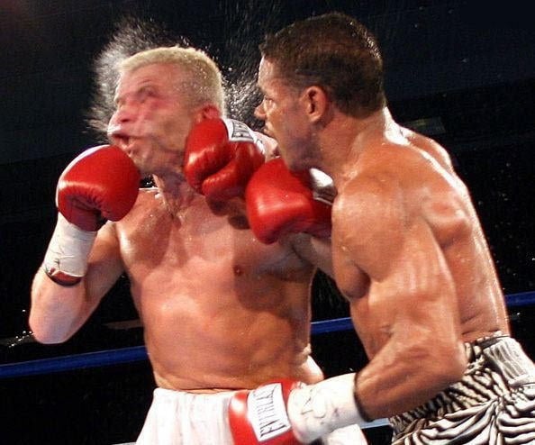
Poster Communications: Boxing head-guards do not prevent Cerebral Trauma
M. R. Graham1,2, T. Myers1, N. E. Thomas4, B. Davies2, S. M. Cooper5, P. J. Evans3, J. S. Baker6
1. PESS, Newman University College, Birmingham, United Kingdom. 2. HESAS, Glamorgan University, Cardiff, United Kingdom. 3. Department of Endocrinology, Royal Gwent Hospital, Newport, United Kingdom. 4. Centre for Child Research, Swansea University, Swansea, United Kingdom. 5. Cardiff School of Sport, University of Wales Institute Cardiff, Cardiff, United Kingdom. 6. Health & Exercise Science Research Unit, University of the West of Scotland, Hamilton, Scotland, United Kingdom.
View other abstracts by:
Where applicable, experiments conform with Society ethical requirements.