
Muscle hypertrophy is an increase in myofibre size requiring net protein synthesis and the activation and subsequent fusion of satellite cells with the existing myofibres. Anabolic agents act to stimulate these processes. Testosterone is a known anabolic agent and has been shown in human skeletal muscle to invoke dose response increases in muscle fibre size and satellite cell number (Sinha-Hikim et al. 2003). The C2C12 mouse muscle cell line has been widely used to study the signalling pathways involved in insulin like growth factor I-induced hypertrophy (Rommel et al. 2001). The purpose of the present study was to determine the suitability of this cell line as an in vitro model for studying the hypertrophic response to androgens, by examining changes in myotube size in response to increasing concentrations of testosterone. Murine C2C12 myoblasts were cultured in a humidified incubator at 37°C and 5% CO2, in growth medium (GM) containing Dulbecco’s Modified Eagle’s Medium (DMEM, Gibco, UK), 1000 g/l glucose and 10% fetal calf serum (FCS). Cells were plated onto 35mm tissue culture dishes at 4×10⁵ cells per dish. After 24 h the cells reached confluency. GM was replaced with differentiation medium (DM, DMEM + 2% horse serum) or serum-free medium (SF) with and without testosterone for 6 days. Media were refreshed every 2 days. The testosterone concentrations were 1nM, 10nM (physiological), 100nM and 1μM. Cells were subsequently fixed in 100% ice cold methanol and digital images obained using a phase contrast microscope (20X). Myotube diameters were quantified according to the method described by Rommel et al. (2001). A total of 50 myotubes were chosen per treatment group. The average diameter per myotube was calculated as the mean of 10 measurements taken at regular intervals along the length of the myotube. The data show that in DM C2C12 myotubes hypertrophy in response to physiological and supraphysiological doses of testosterone (p<0.05, ANOVA). In SF medium, myotubes were significantly (p<0.05) smaller than those in DM at all concentrations. The myotubes in SF medium were also responsive to testosterone (p<0.05), albeit to a lesser extent. The mean increase in myotube size from control to the 1µM testosterone concentration was 36% in DM and 17% in SF medium. This indicates that factors in addition to testosterone are regulating myotube size in DM. Taken together, the data suggest that C2C12 cells may provide an appropriate model for studying androgen-induced mechanisms of muscle hypertophy.
University College London 2006 (2006) Proc Physiol Soc 3, PC125
Poster Communications: C2C12 muscle cell line as a model to study androgen-induced myofibre hypertrophy
Tomasz George1, Stephen Harridge1, Cristiana Velloso1
1. Applied Biomedical Research, King's College London, London, United Kingdom.
View other abstracts by:
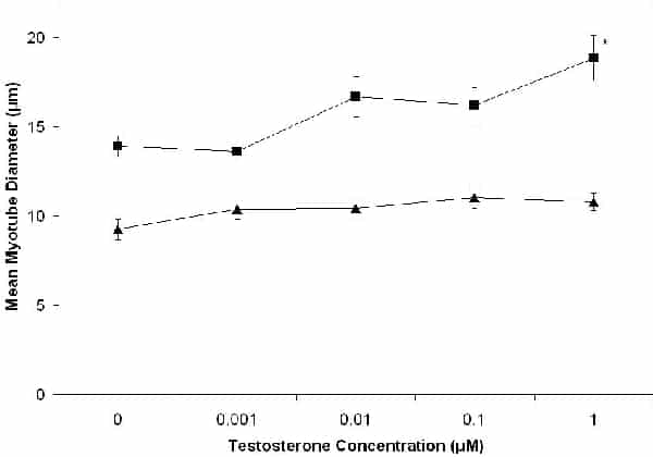
Figure 1. Myotube size in response to increasing concentrations of testosterone in either serum-free medium (triangles) or differentiation medium (squares). * Indicates significant difference from 0 μm testosterone (p<0.05 post-hoc t tests corrected for multiple comparisons). Data points are mean ± sem n=50.
Where applicable, experiments conform with Society ethical requirements.