
H441 cells express an amiloride-sensitive Na+ conductance (GNa) similar to that associated with co-expression of the epithelial Na+ channel (ENaC) α, β and γ subunits, and regulated increases in GNa seem to underlie the cAMP-evoked stimulation of Na+ transport seen in these cells (Clunes et al., 2004). However, as well as increasing [Ca2+]i, thapsigargin (1 μM) also increases the short circuit current recorded from these cells (ΔISC = 24.3 ± 2.4 μA cm2, n = 6, P < 0.01), suggesting that Na+ transport is also controlled via [Ca2+]i. This was unexpected as Ca2+ is thought to inhibit ENaC (Ishikawa et al., 1998) and so, to establish the physiological basis of this response, we explored the effects of thapsigargin (1 μM) upon the membrane currents recorded from groups of 2-5 cells (Clunes et al., 2004). Thapsigargin consistently evoked Ca2+-dependent membrane currents (Fig 1A) and the current recorded from thapsigargin-treated cells reversed at a potential close to EK (Fig 1B). This Ca2+-mobilising agent had thus increased GK and further analysis showed that this was associated with a hyperpolarization of Vm (control: -50 ± 6 mV; thapsigargin: -78 ± 1 mV, P < 0.01). We also studied the effects of thapsigargin upon GNa by characterising amiloride-sensitive currents in cells treated with clotrimazole (1 μM) which was shown to block the thapsigargin-evoked increase in GK (Clunes et al., 2005). These experiments confirmed the expression of a selective Na+ conductance and showed that the magnitude of this conductance increased slightly during exposure to thapsigargin (control: 157 ± 16 pS cell-1; thapsigargin: 267 ± 24 pS cell-1, n = 7, P < 0.02) a result that contrasts with earlier work (Ishikawa et al., 1998). Thapsigargin may thus stimulate Na+ transport by (i) hyperpolarising Vm and increasing the driving force for Na+ entry and (ii) increasing GNa by an unknown mechanism.
King's College London (2005) J Physiol 565P, C10
Communications: Ca2+-evoked membrane currents in a cell line (H441) derived from the human bronchial epithelium
McNeill, Richard P; Clunes, Mark T; Chambers, Lucy A; Wilson, Stuart M;
1. TICH, University of Dundee, Dundee, Tayside, United Kingdom.
View other abstracts by:
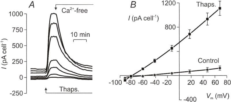
Fig. 1. Dexamethasone-treated (0.2 μM) H441 cells grown on glass coverslips were held under voltage clamp in the perforated patch recording configuration. The membrane currents were evoked by driving ψ*bsup*+ s.e.m. n = 9) in control and thapsigargin-treated cells.
Where applicable, experiments conform with Society ethical requirements.