
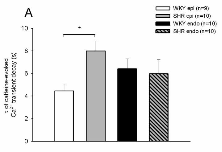
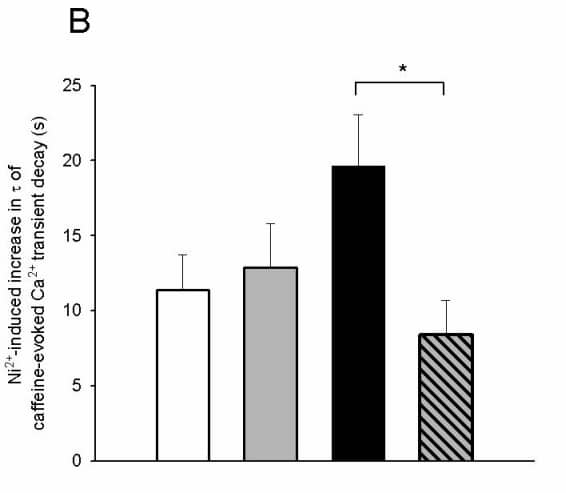
Pressure overload-induced hypertrophy produces regional changes in cardiac cell structure and function. We have therefore investigated the changes in Ca2+ handing that occur in myocytes from the left ventricular sub-epicardium (EPI) and sub-endocardium (ENDO) of 20 week old spontaneously hypertensive rats (SHR) compared with normotensive age-matched Wistar-Kyoto (WKY) control rats.Rats were killed humanely (Schedule 1 method) and ventricular myocytes isolated by enzymatic dispersion. Cells were superfused with Tyrode solution at 22-24 °C and electrically stimulated via extracellular electrodes. Intracellular Ca2+ was monitored using fura-2.Following a train of 1 Hz conditioning stimuli, rapid application of caffeine (20 mM) was used to release Ca2+ from the sarcoplasmic reticulum (SR). The amplitude of the electrically stimulated and caffeine-induced Ca2+ transients was significantly bigger (P<0.02 and P<0.05, respectively; unpaired t-tests) in SHR EPI cells than in WKY EPI cells and fractional release remained unchanged. In contrast, there was no significant difference between the amplitude of the electrically stimulated or caffeine-induced Ca2+ transients, or fractional release in ENDO cells from SHR and WKY hearts. The time constant of decay of the caffeine-induced Ca2+ transient (τ; which reflects sarcolemmal Ca2+ extrusion) was significantly greater (P=0.005) in SHR EPI than in WKY EPI cells, but was not significantly different in SHR and WKY ENDO cells (Fig. 1A). Simultaneous application of caffeine and 10 mM Ni2+ (to inhibit Ca2+ extrusion via Na/Ca exchange; NCX) increased τ in all cell types (Fig. 1B). In EPI cells, the Ni2+-induced increase in τ was not significantly different between SHR and WKY cells (Fig. 1B). However, the increase in τ was significantly smaller (P=0.015) in SHR ENDO than WKY ENDO cells (Fig. 1B). Fig. 1. A. τ of decline of the caffeine-induced Ca2+ transient in WKY and SHR EPI and ENDO cells. B. Ni2+-induced increase in τ in each cell type. * indicates statistical significance.Thus the slower decline of the caffeine-induced Ca2+ transient in SHR EPI cells in the absence of Ni2+ does not appear to be due to lower NCX activity, suggesting that another Ca2+ extrusion pathway may be down-regulated in these cells. In contrast, τ was similar in SHR and WKY ENDO cells in the absence of Ni2+; the smaller effect of Ni2+ in SHR ENDO cells suggests less NCX-mediated Ca2+ extrusion in these cells, and hence that other mechanisms may be up-regulated to compensate for this apparent decrease in NCX activity.
University of Glasgow (2004) J Physiol 557P, C1
Communications: Ca2+ handling in ventricular myocytes isolated from the sub-epicardium and sub-endocardium of the spontaneously hypertensive rat heart.
J. Naz, M.R. Fowler, S.M. Harrison and C.H. Orchard
Biomedical Sciences, University of Leeds, Leeds, West Yorkshire, UK
View other abstracts by:
Where applicable, experiments conform with Society ethical requirements.