
Human skeletal muscle has the potential to regenerate completely after injury induced under controlled experimental conditions. Examples of models suitable for humans include voluntary eccentric contractions or contractions induced by neuromuscular electrical stimulation (NMES). The major difference between these two models appears to be the proportion of muscle fibres damaged to the point of necrosis, with voluntary contractions rarely leading to muscle fibre necrosis in prime mover muscles (Crameri et al., 2007). In studies where NMES has been employed, the proportion of necrotic fibres appears to range from approximately 15-40% (Crameri et al., 2007; Mackey et al., 2016; Karlsen et al., 2020). While the events inside the myofibers as they undergo necrosis, followed closely by satellite cell-mediated myogenesis, have been described in detail (Mackey & Kjaer, 2017), much less is known about the involvement of the key mononuclear cell populations such as fibroblasts, immune cells and vessel associated cells. Furthermore, while the adaptation of the connective tissue structures surrounding the myofibres throughout the degeneration and regeneration processes is gaining interest, the role of individual muscle matrix components and their spatial interaction during repair is poorly understood – and may provide insight into the optimal timing of rest vs. return to activity after muscle injury. This not only relates to restoration of the insults to the muscle mid-portion, but also to the interface between the muscle fibres and the tendon – the myotendinous junction (MTJ). In general, it appears that the muscle connective tissue takes longer to recover after contraction induced damage than the muscle fibres themselves, which may explain the high rate or re-injury recorded after an initial strain injury. While progress is being made in determining the composition of the intact human MTJ (Karlsen et al., 2022), many questions remain regarding the protein and cellular composition with injury and during repair.
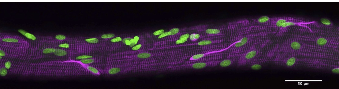
Figure shows an almost fully regenerated human skeletal muscle fibre (desmin, magenta; nuclei, green). Note the three desmin-positive satellite cells on the myofibre surface.
Biomedical Basis of Elite Performance 2022 (University of Nottingham, UK) (2022) Proc Physiol Soc 49, SA10
Research Symposium: Cell and extracellular matrix interactions of skeletal muscle fibres
Abigail L. Mackey1,2,3
1 Institute of Sports Medicine Copenhagen, Department of Orthopaedic Surgery M, Copenhagen University Hospital - Bispebjerg and Frederiksberg 2 Xlab, Department of Biomedical Sciences, Faculty of Health and Medical Sciences, University of Copenhagen 3 Centre for Healthy Aging, Department of Clinical Medicine, Faculty of Health and Medical Sciences, University of Copenhagen
View other abstracts by:
Where applicable, experiments conform with Society ethical requirements.