
Interleukin-1 (IL-1) is the prototypical pro-inflammatory cytokine, and is implicated in the pathogenesis of acute and chronic neurodegenerative diseases (Allan et al., 2005). Classical IL-1 signalling does not fully account for the effects of IL-1 observed in the literature (Touzani et al., 2002). The pro-forms of the IL-1 family agonists, IL-1α and β, contain a confirmed and a putative nuclear localisation sequence respectively, and evidence suggests that IL-1α has intranuclear actions in IL-1 expressing cells (Wessendorf et al., 1993; Werman et al., 2004). Microglia are an early source of IL-1 in the injured central nervous system (CNS, Pearson et al., 1999). Therefore we tested the hypothesis that IL-1 isoforms localise to microglial nuclei following inflammatory stimuli. Murine primary microglia and a murine microglial cell line (BV-2) were treated with bacterial lipopolysaccharide (LPS) to induce IL-1 expression, and IL-1 subcellular localisation was characterised by immunocytochemistry. The proportion of cells containing intranuclear IL-1 was quantified by blind counting of IL-1 immunostained cells. LPS induced pro-IL-1α and β localised to the cytosol and nucleus of both primary microglia and BV-2 cells. Nuclear localisation was more pronounced for IL-1α than for IL-1β. IL-1 subcellular localisation in BV-2 cells was cell density dependent, being predominantly cytosolic at high density, and both nuclear and cytosolic at low density (Figure 1). Cytosolic localisation of IL-1 in BV-2 cells cultured at low density was induced by co-culture with a confluent HEK cell monolayer, indicating that IL-1 localisation may be regulated by cell contact. Thus IL-1α and β localise to microglial nuclei in a regulated fashion, and may therefore have intra-nuclear actions in microglia regulating CNS inflammatory responses.
Life Sciences 2007 (2007) Proc Life Sciences, PC121
Poster Communications: Cell density regulates nuclear localisation of interleukin-1α and β in murine microglia
N. M. Luheshi1, N. J. Rothwell1, D. Brough1
1. Faculty of Life Sciences, University of Manchester, Manchester, United Kingdom.
View other abstracts by:
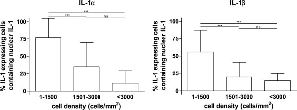
Figure 1: Cell density dependent nuclear localisation of IL-1α and β in BV-2 cells. BV-2 cells seeded at various densities were treated with 1μg/ml LPS for 6h fixed immunostained for IL-1 α or β and nuclei labelled with DAPI. Random fields of view were chosen by focusing on the DAPI stain and widefield images were captured using a 40x objective. Local cell density was assessed by counting DAPI stained nuclei and the proportion of IL-1 expressing cells containing nuclear IL-1 was counted in each field of view. The data shown are mean ± SD from three independent experiments. *** p<0.001 Kruskall Wallis with post hoc Dunn’s multiple comparison test.
Where applicable, experiments conform with Society ethical requirements.