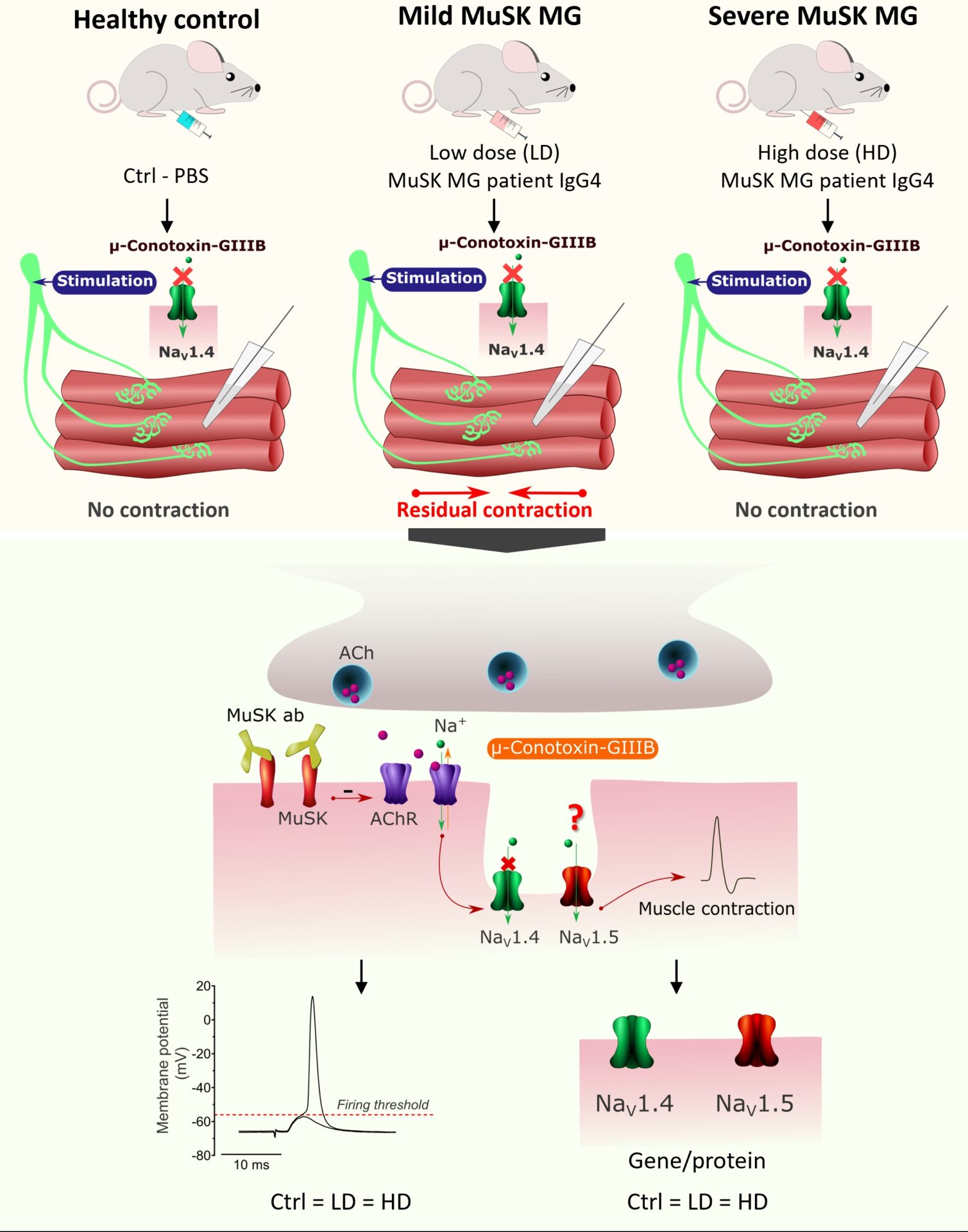
Muscle-specific kinase myasthenia gravis (MuSK MG) is an autoimmune neuromuscular disorder. It is caused by autoantibodies against MuSK, a key postsynaptic membrane molecule involved in acetylcholine receptor clustering at the neuromuscular junction (NMJ). MuSK MG patients have fluctuating, fatigable weakness. The disease severity can vary greatly between patients, in spite of comparable autoantibody levels. One explanation for inter-patient and inter-muscle variability in sensitivity might be variations in compensatory muscle responses. Previously, we developed a passive transfer mouse model for MuSK MG, using purified patient IgG4. In our ex vivo experiments we observed that nerve stimulation-evoked contraction of muscles from mild MuSK MG mice appeared partly insensitive to μ‑Conotoxin-GIIIB. This blocker of NaV1.4 voltage-gated sodium channels normally completely eliminates muscle contraction. We hypothesized that changes in NaV channel expression profile, possibly compensatory expression of (μ-Conotoxin-GIIIB insensitive) NaV1.5 cardiac-type channels, might lower the muscle fibre’s firing threshold and improve neuromuscular synaptic transmission in mild MuSK MG.
We performed passive transfer in immuno-deficient NOD.CB17-Prkdcscid/J (Nod/scid) mice, using ‘high’(n=12), ‘intermediate’ (n=3) and ‘low’ (n=12) dosing regiments of purified MuSK MG patient IgG4. Next, we assessed myasthenia levels, μ-Conotoxin-GIIIB resistance and firing thresholds. Ex vivo analysis of myasthenic NMJs was performed by evaluation of miniature endplate potentials and endplate potentials in the presence of μ-Conotoxin-GIIIB in diaphragm-nerve preparations. To detect changes in NaV1.4 and NaV1.5 channels expression profile we performed qPCR and immunostaining in muscle tissue. Data is presented as mean ± S.E.M., with n representing the number of animals per group. Statistical analysis was performed using multiple unpaired t-tests or one-way ANOVA with Tukey’s post hoc tests. All procedures involving the use of laboratory animals were performed in accordance with Dutch law and Leiden University guidelines.
High- and intermediate-dosed mice showed severe, progressive myasthenia, not detected in low-dosed animals. However, during electrophysiological analysis of diaphragm NMJs we found equally reduced amplitudes and frequencies of miniature endplate-potentials (MEPPs), and equal reductions of endplate potentials (EPPs) amplitudes (all by~40-50%, as compared to the PBS-control), probably associated with direct exposure to MuSK MG patient IgG4 via the intraperitoneal injection route. Notably, the diaphragms from low-dosed mice (n=12) showed a much higher degree of μ-Conotoxin-GIIIB resistance according to the scale for contraction responses when compared with PBS mice (n=7) or high-dosed mice (n=12). However, Scn5a, the gene encoding NaV1.5, was not upregulated in muscle tissue of these mice (PBS mice: n=4, low-dosed mice n=6, high-dosed mice n=6; p > 0.05). Furthermore, no reduction of firing thresholds or expression of histologically detectable NaV1.5 channels was found (PBS mice: 8.61 ± 0.11 mV (n=4); low-dosed mice: 8.60 ± 0.28 mV (n=6); high-dosed mice 8.69 ± 0.22 mV (n=6)). Thus, we observed NaV1.4-independent muscle contraction ex vivo, preferentially in mice experiencing subclinical MuSK MG, which is unlikely associated with upregulation of NaV1.5. It remains to be established which other NaV channels or factors are responsible for the observed μ-Conotoxin-GIIIB insensitivity and if the change in NaV repertoire is compensatory beneficial.