The sarcoplasmic reticulum acts as the main intracellular store of Ca used to activate contraction within cardiac myocytes. Mechanisms which increase the Ca content of the SR are positively inotropic, although they can also lead to after-depolarisations thought to be involved in arrhythmogenesis.
Electrical remodelling in the atrium occurs in response to prolonged rapid rates of stimulation and leads to an abbreviation of the action potential duration and to self perpetuation of atrial arrhythmias. A reduction of the L-type Ca current has been shown in humans with atrial fibrillation. Calcium overload has also been shown in electrically remodelled atrial cells.
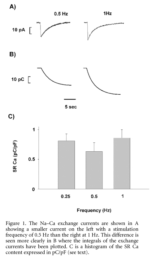
The aim of the current experiments was to determine whether changing the rate of stimulation alters the Ca load of the SR. Cells were isolated from humanely killed male Wistar rats (200-250 g) by a collagenase and protease digestion technique. Cells were voltage clamped using the perforated patch technique and stimulated at rates of 0.25, 0.5 and 1 Hz at 37 °C. Fluorescence measurements were performed using fluo-3. Caffeine (10 mM) was applied to discharge the SR of Ca and the resulting Na-Ca exchange current (Fig. 1A) was integrated (Fig. 1B) to determine SR Ca content.
Results were obtained for n = 8 myocytes. SR Ca content, expressed as the integral relative to cell capacitance in pC/pF, was found to be on average 0.81 ± 0.15 at 0.25 Hz, 0.63 ± 0.12 at 0.5 Hz and 0.86 ± 0.14 at 1 Hz (Fig. 1C). Using paired t tests, a statistically significant difference was found comparing 0.5 Hz with 0.25 Hz (P < 0.05) and 0.5 Hz with 1 Hz (P < 0.05), but not comparing 0.25 Hz with 1 Hz. The changes seen in the SR Ca content were mirrored in the systolic Ca transients which were larger at 0.25 and 1 Hz compared with 0.5 Hz. It remains to be seen whether these changes can be accounted for by differences in the L-type Ca current and what changes occur at higher rates.
All procedures accord with current UK legislation.