
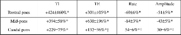
Following transection of rostral pontile regions in anaesthetised cats in vivo, Lumsden (1923) described apneusis, whereas a complete removal of the pons resulted in gasp-like discharges. Due to confounding problems relating to anaesthesia and haemorrhage, as well as not knowing where exactly the lesions were made, we refined these experiments to avoid effects of anaesthesia/poor brainstem perfusion and reconstructed brainstem transections precisely. Wistar rats (male; 65-85g) were anaesthetised deeply with halothane until they became unresponsive to noxious pinching of the tail. They were decerebrated at the pre-collicular level and perfused arterially (Paton, 1996). Recordings of the phrenic (PN), central vagal (X) and abdominal (AB) motor nerves were made during transverse sectioning of the brainstem. In the in situ preparation, eupnoea was characterized by ‘ramp-like’ inspiratory activity of the PN, presence of post-inspiratory activity (PI) in recordings of X and little or no activity in the AB. Three distinct pontine levels were transected as confirmed histologically in sagittal sections in separate rats: (1) rostral pons (5.1-5.7 mm rostral to calamus scriptorius, CS, n=5); (2) mid-pons (4.4-4.8 mm rostral to CS; n=3); (3) caudal pons or immediately rostral to the facial nucleus (i.e. ponto-medullary junction; 4.0 mm rostral to CS; n=8). Transection at all 3 levels resulted in apneusis, increased expiratory time and loss of PI (Table 1). Transection of the rostral pons resulted in apnoea in 2 of 5 preparations but the rhythm could be reinstated by either a subsequent transection or increasing respiratory drive (7.5% CO2). Changes in PN parameters did not differ between rostral and mid-pontile transections (P>0.05, t test) compared to control. However, caudal pons transection produced a smaller depression of amplitude and rate of PN compared to the more rostral cuts (Table 1). In addition, tonic expiratory activity in the AB nerve occurred after transection of the caudal pons. Consistent with Lumsden’s (1923) observations we conclude that the pons is essential for the generation of the normal eupnoeic motor discharge in situ. The apnoea and depressed respiratory motor outputs evoked after disconnection of either rostral or mid-pons is caused, in part, by descending inhibitory drives generated in the caudal pons, perhaps by the A5 cell group. Finally, the pons seems to depress AB expiratory activity, as it is not present until its removal.
University College London 2006 (2006) Proc Physiol Soc 3, PC87
Poster Communications: Changes in the pattern of spinal and cranial respiratory motor outflows after micro, transverse sectioning of the pons in situ
Ana P.L. Abdala1, Jeffrey C. Smith2, Ilya A. Rybak3, Julian F.R. Paton1
1. Department of Physiology, Bristol Heart Institute, School of Medical Sciences, University of Bristol, Bristol, Avon, United Kingdom. 2. Cellular & Systems Neurobiology Section, Laboratory of Neural Control, National Institute of Neurological Disorders & Stroke, National Institute of Health, Bethesda, MD, USA. 3. School of Biomedical Engineering, Science and Health Systems, Drexel University, Philadelphia, PA, USA.
View other abstracts by:
Where applicable, experiments conform with Society ethical requirements.