
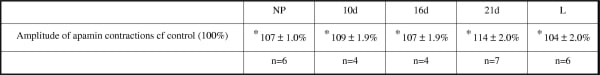
SK channels are small conductance Ca2+ – activated K+ channels which may play an important in maintaining uterine quiescence, as the outward movement of potassium ions serve to re-polarise or hyperpolarize the myometrium. As the relative quiescence of the myometrium changes throughout gestation, the aim of this study is to determine the expression of all three isoforms of SK channels (SK1-3) and their contribution to normal rhythmic contractility throughout pregnancy in the rat. Whole uterine horns were dissected from nonpregnant (NP) Wistar rats and at different stages of pregnancy:- 10d, 16d, 21d, labouring (L; term 22d). The expression of SK 1-3 was characterised at each stage using RT-PCR, immunohistochemistry on grouped samples in tissue microarrays, and quantitative western blotting. Measurement of spontaneous phasic contractions were made on strips of longitudinal myometrium at each stage and the role of SK channels was determined by comparing contractions over a 20 min control period to those during a 20 min application of apamin (100nM), a constituent of bee venom that inhibits all three isoforms of SK channels [1]. Consistent expression of all SK isoform transcripts was seen in all samples. In NP and at all stages of pregnancy, clear staining of SK 1-3 was seen in the myometrial cell layer. No staining was seen in epithelial cells. As the immunostaining of SK2 and SK3 appeared to change in density on the tissue microarrays in late pregnancy, expression of SK2 and SK3 was quantified by western blotting. Both proteins were clearly seen in all samples. There was no change in the expression of SK3 throughout pregnancy, but compared to β actin controls, expression of SK2 protein was significantly decreased in 16d and 21d compared with 10d and NP rats, and then increased in L tissue. Apamin had no effect on the frequency of contractions but significantly increased the amplitude of spontaneous myometrial contractions in all stages of rat pregnancy (table 1). These data clearly show that SK 1-3 channels are expressed in rat myometrium and limit spontaneous contraction in native, myometrial strips. SK channels appear to inhibit uterine contractions to a lesser extent than in the bladder, where the effect of 100nM apamin on contaction amplitude was ten fold greater than in the present study [1]. These data also show that SK2 is downregulated during late pregnancy which is in agreement with Mazzone & Buxton who found that SK2 is downregulated in human myometrium at term, [2]. This gestation-dependant regulation of SK2, merits further study. Reference 1 : Herrera G & Neson M (2002), J Physiol. 541, 483 – 492 Reference 2 : Mazzone J & Buxton IL (2003), Proc. West. Pharmacol. Soc. 46, 74 – 77
University of Edinburgh (2007) Proc Physiol Soc 6, PC5
Poster Communications: Characterisation and functional significance of SK channels in the pregnant rat uterus.
K. Noble1, R. Floyd1, A. Mobasheri2, S. Wray1
1. Physiology, University of Liverpool, Liverpool, United Kingdom. 2. Veterinary Medicine & Science, University of Nottingham, Nottingham, United Kingdom.
View other abstracts by:
Where applicable, experiments conform with Society ethical requirements.