
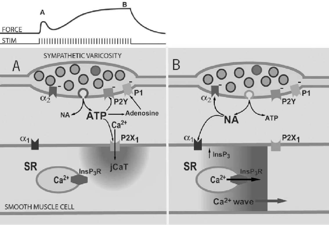
Small mesenteric arteries contribute significantly to the regulation of blood flow and to total peripheral resistance in the body. In these arteries, control of smooth muscle contraction by the sympathetic nervous system is of major importance. This control is complex, involving multiple neurotransmitters and receptors, as well as complex patterns of nerve fibre activity. For example, three different sympathetic neurotransmitters (ATP, NA and neuropeptide Y) are released at sympathetic neuroeffector junctions, and the effects of these different neurotransmitters are thought to be synergistic in activating smooth muscle Ca2+ signaling and contraction. Furthermore, the neural release of ATP and NA varies with the pattern of nerve fibre activity. In the face of such complexity, simple bath application of one neurotransmitter (e.g. NA) to isolated smooth muscle cells, as classically done, cannot be expected to reveal the events involved in the physiological control of arterial contraction by the sympathetic nervous system. Therefore, we and others have developed methods recently to visualize Ca2+ signaling in smooth muscle cells of intact arteries, during sympathetic nerve stimulation and development of force (isometric). Confocal myographs are an adaptation of classical wire myographs used to study contractions of blood vessels, which permit mounting the artery such that confocal imaging can be performed and perivascular nerves stimulated. Use of long working distance, high numerical aperture water objective lenses is necessary. As known from previous work, neurogenic contractions of small arteries consist of an early, relatively small and transient component that is activated mainly by ATP acting on P2X receptors (marked A on the force record Fig.1), and a later, larger, slowly developing component that is activated mainly by NA acting on alpha-1 adrenoceptors (marked B). Recent confocal Ca2+ imaging (fluo4 fluorescence) with the confocal myograph reveals that early during a train of nerve fibre action potentials, smooth muscle contraction is activated mainly by post-junctional Ca2+ transients (jCaTs) (Lamont & Wier, 2002) induced by neurally released ATP. JCaTs are localized to the post-junctional region, and arise from Ca2+ that has entered via P2X receptors. At this time, sympathetic varicosities (neuroeffector junctions) may release mainly small vesicles that contain a relatively high concentration of ATP (viz. the relatively few big quanta proposed by Stjarne, 2001) (schematic A). Later during a train of nerve fibre action potentials, we found that jCaTs are rare (Lamont, Vainorius & Wier, 2003), and contraction is activated by Ca2+ waves that arise from sarcoplasmic reticulum (SR). These Ca waves appear identical to those activated by bath-applied NA and arise from Ca release from SR that is activated by inositol tris-phosphate. At this time, sympathetic varicosities may release small synaptic vesicles that contain a relatively high concentration of NA (schematic B). Notable differences in Ca2+ signaling during neurogenic contractions and those elicited by bath-applied neurotransmitters are: 1) the spike and plateau pattern of intracellular Ca2+ seen in isolated smooth muscle cells after application of NA is never seen in the smooth muscle cells of intact arteries during nerve stimulation, and 2) neurally released ATP does not elicit Ca2+ waves in smooth muscle cells of intact arteries, as does bath-applied ATP in isolated smooth muscle cells. We suggest that these differences in Ca2+ signaling arise from the very different patterns in spatio-temporal patterns of transmitter concentration and receptor activation that occur with neural stimulation as opposed to bath-application of ATP and NA. The use of intact small arteries and the confocal myograph thus provides a useful new tool for revealing mechanisms involved in control of vascular resistance by the sympathetic nervous system.
University of Glasgow (2004) J Physiol 557P, SA18
Research Symposium: Confocal Ca Imaging in Intact Small Arteries Reveals Smooth Muscle Ca2+Transients Attributable to Neurally Released ATP and NA
W.G. Wier and C. Lamont
Physiology, University of Maryland, Baltimore, Baltimore, MD, USA
View other abstracts by:
Where applicable, experiments conform with Society ethical requirements.