Direct vagal stimulation studies reveal significant decreases with postnatal age in the influence of the parasympathetic control of heart rate (Quigley et al. 1996). Accordingly, the electrical properties of autonomic ganglion neurones from neonatal rats appear to be different from those of mature, adult rats (Hogg et al. 2001).
This research aims to determine the diversity of electrophysiological properties of intracardiac ganglion neurons in a wholemount preparation of intracardiac ganglia from adult (²ge³ 6 weeks) and neonatal (2Ð5 days) Wistar rats (killed humanely in accordance with the guidelines of the University of Queensland Animal Experimentation Ethics Committee). Intracellular recordings were made using conventional sharp glass microelectrodes and the preparation was superfused with a bicarbonate-buffered physiological salt solution maintained at 37 °C.
In adult neurones, the average resting membrane potential (Em) was -53.6 ± 1.6 mV (mean ± S.E.M., n = 27) and the input resistance (Rin) was 97.5 ± 11.5 MΩ. The Em and Rin for neurones from neonatal rats was -49.5 ± 1.8 mV and 80.7 ± 17.6 MΩ (n = 5), which is similar to that of adult ganglion neurones.
In agreement with a previous report (Selyanko, 1992), at least two neurone types were identified in ganglia from adult rats according to the duration of the after-hyperpolarization (duration to 50 % recovery, AHP50); neurones which display a brief AHP50 (< 30 ms) following somatic action potentials (20.9 ± 2.0 ms, n = 10) and long duration AHP50 > 50 ms (68.1 ± 5.1 ms, n = 6). This difference was significant (P < 0.001, unpaired t test). There was, however, no significant difference in the AHP amplitude between these groups (-14.0 ± 1.3 and -15.6 ± 1.5 mV) for brief and long duration AHP50 groups, respectively.
In neonatal rat intracardiac neurones, the AHP was shallow (-5.3 ± 1.6 mV, n = 3) and of only short duration (AHP50 23.4 ± 8.3 ms).
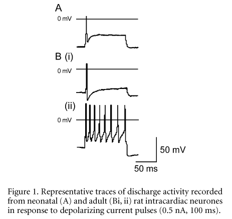
Neonatal neurones (n = 5) discharged a single action potential in response to long duration depolarizing currents, whereas adult neurones exhibited a diversity of responses from fast adapting to sustained discharge (see Fig. 1).
A Travel award from The Wellcome Trust to A.A.H. and NHMRC grants to D.J.A. supported this work.
All procedures accord with current local guidelines.