
Mechanosensory nerve terminals, including those of the muscle spindle, possess an autogenic glutamatergic modulatory mechanism, mediated by the non-canonical metabotropic action of a phospholipase D-linked receptor1. We have previously reported to the Society our evidence that the receptor is the kainate receptor, GluK22. We have now compared the responses of muscle spindles from GluK2-disrupted (GluK2-D) and wild type littermate (WT) mice, each exposed to a repeated battery of stretch profiles, to test the effects of periods of complete unloading to minimise endogenous glutamate release and exogenous glutamate application (Fig 1). Here we report the actions of exogenous glutamate on responses during the hold phase of ramp stretches. Experiments were performed under relevant national animal experimentation licences, and local ethical committee approvals. Adult GluK2-D mice had the Grik2 gene disrupted by insertion of a Neo-cassette into MD2 (from Prof Christophe Mulle, Univ. of Bordeaux)3. Soleus nerve-muscle preparations were removed after humane killing (Schedule 1, Animals (Scientific Procedures) Act, 1986). Afferent firing in response to stretches was recorded as the electroneurogram using a suction electrode. The numbers of action potentials (APs) fired during the hold phases of the series of stretch-hold-release (ramp) profiles were counted and compared between genotypes in the absence and then the presence of glutamate added exogenously at 100 µM and 1 mM. Data are expressed as mean ± SEM, from ‘n’ muscles and ‘m’ mice. Statistical analysis was carried out by repeated measures ANOVA. When 100 µM glutamate was increased to 1 mM, GluK2-D mouse spindles displayed a very modest reduction in firing (342 ± 3 vs 299 ± 3 APs; P < 0.001; n = 5, m = 9). In contrast, spindles in WT mice showed one of two responses to the change: either a marked increase in firing (440 ± 5 vs 501 ± 6 APs; P < 0.001; n = 4, m = 2), or a very marked decrease in firing (139 ± 3 vs 52 ± 2 APs; P < 0.001; n = 3, m = 2). At present we can only speculate about the mechanisms of decreased firing in some WT mice. We suggest it may reflect receptor desensitisation, since kainare receptors are very prone to desensitisation4, or excitotoxicity from prolonged exposure to high concentrations of glutamate. We conclude that the absence of an excitatory action of glutamate in the GluK2-D mice is due to the functional disruption of GluK2 receptors, indicating an important role for GluK2 in spindle peripheral terminal modulation.
Physiology 2019 (Aberdeen, UK) (2019) Proc Physiol Soc 43, PC214
Poster Communications: Disruption of the actions of exogenous glutamate on the stretch-evoked responses of muscle spindles in GluK2-deficient mice
R. W. Banks3, C. Mulle2, G. S. Bewick1
1. Institute of Medical Sciences, University of Aberdeen, Aberdeen, Scotland, United Kingdom. 2. The Neuroscience Institute at Bordeaux, Universite de Bordeaux, Bordeaux, France. 3. Department of Biosciences, University of Durham, Durham, United Kingdom.
View other abstracts by:
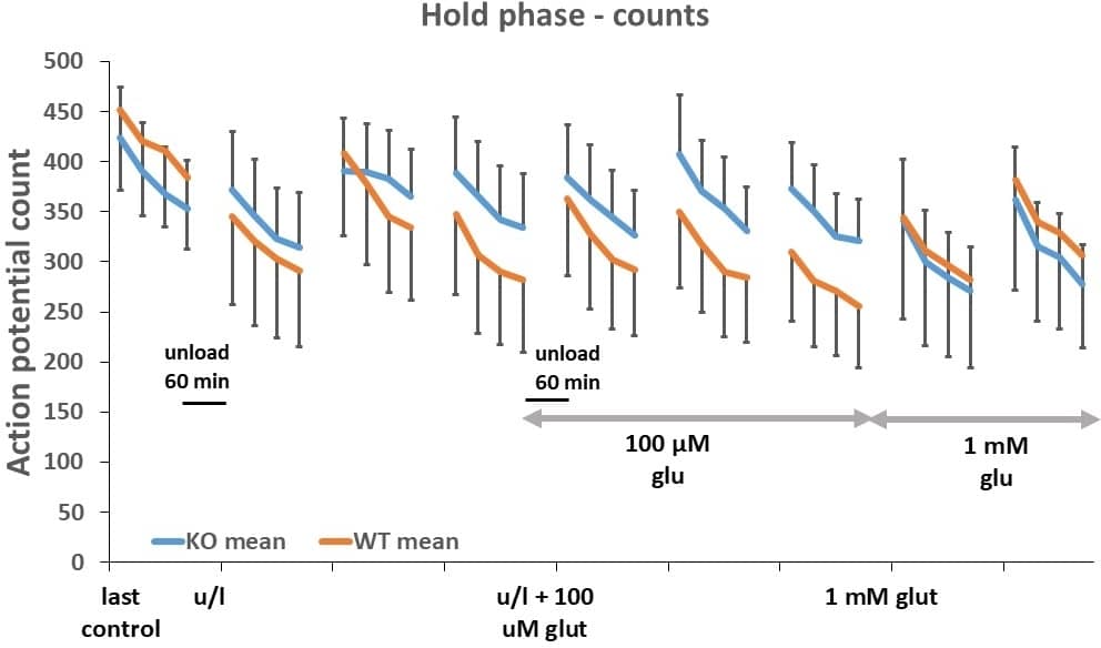
Muscle spindle action potential counts during hold phase of ramp stretch in GluK2-D and WT mice during series of unloading and exogenous glutamate applications. Increasing glutamate from 0.1 to 1 mM caused mild inhibition of firing in GluK2-D mice, but increased mean firing in WT. Mean � SEM, n = 5 (GluK2-D) and 7 (WT).
Where applicable, experiments conform with Society ethical requirements.