
Cardiac output is thought to be the main limiting factor of maximal aerobic exercise in healthy subjects (Coats, 2001). Alterations in interventricular (interV) and intraventricular (intraV) synchrony of the heart produces change in cardiac output during exercise in heart failure patients (Nowark et al.1995). There is however no research into the effect of exercise on ventricular synchrony in healthy subjects where alterations in ventricular timings may represent more favourable haemodynamics for peak exercise. Approval for this study was granted by the East Sussex Local Research Ethics Committee and conducted in accordance with the declaration of Helsinki. Five male subjects (mean±SEM age: 27.8±2.5yrs, mass: 80±1.6kg, BMI: 24.7±0.8 kg/m2) took part in the study. Inclusion required a QRS duration <120ms indicative of normal electrical conduction, as determined by 12-lead ECG. Each subject completed an incremental exercise test on a recumbent exercise bicycle to volitional exhaustion. Echocardiographic readings were recorded at rest and peak exercise using a commercially available system. Images were analysed offline. To assess interV synchrony (IVS), the difference between the LV and RV ejection times was analysed (Ghio et al.2004). Ejection time was defined as the time from q wave to flow assessed using pulsed wave Doppler in the right and left ventricular outflow tracts. An IVS>40ms was considered as dyssynchronus (Ghio et al.2004). To analyse IntraV synchrony pulsed wave tissue Doppler imaging (TDI) was performed using apical two and four chamber views at the level of the mitral annulus allowing analysis of longitudinal function of the anterior, inferior, septal and lateral walls of the left ventricle. All the possible differences between the peak contraction time (PCT) of the four basal segments were calculated. IntraV dyssynchrony was considered to be present if absolute differences between any two segments was greater than 1 standard deviation of the regional PCT (Ghio, 2004). Statistical analysis was achieved using paired t-tests. All subjects displayed interV synchrony at rest and at peak exercise. The PCT for each of the LV segments at exercise were significantly greater when compared to rest (P<0.01). No significant effect of exercise on interV synchrony was observed (P=0.638). A tendency was observed for the lateral/anterior (P=0.097) and the anterior/inferior (P=0.112) synchrony (Table 1) to reverse at peak exercise suggesting that further investigation may reveal an alteration in intraV timings at peak exercise.
King's College London (2005) J Physiol 565P, PC19
Communications: Does ventricular synchrony change with exercise in healthy men?
Mullan, Paul ; Silberbauer, John ; Llyod, Guy ; Carr-White, Gerry ; Brickley, Gary ;
1. Chelsea School, University of Brighton, Eastbourne, East Sussex, United Kingdom. 2. Coronary School Research Centre , Eastbourne District General Hospital, Eastbourne, East Sussex, United Kingdom.
View other abstracts by:

Table 1. IntraV timings (ms) at rest and at peak exercise (mean± SEM)
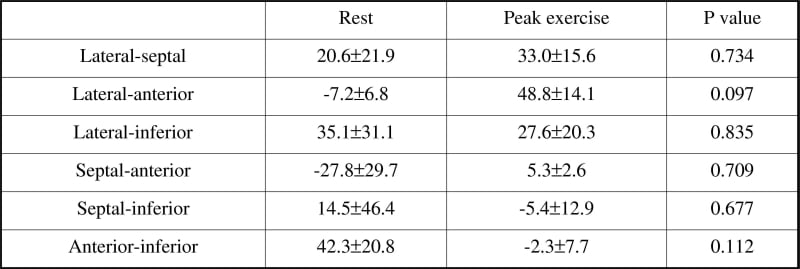
Table 1. IntraV timings (ms) at rest and at peak exercise (mean± SEM)
Where applicable, experiments conform with Society ethical requirements.