
Primary afferent depolarization (PAD) is a phenomenon associated with pre-synaptic inhibition of the effects of primary afferents on their synaptic targets in the spinal cord. PAD is readily evoked by stimulation of other afferents in the same, or closely neighbouring spinal segments. The mechanisms underlying this have been extensively studied (e.g. Rudomin & Schmidt, 1999; Willis, 1999). With electrical stimulation, dorsal root potentials (DRPs which are often taken as a sign of PAD) are also evoked in the roots of more distant spinal segments (e.g. Lidierth & Wall, 2001). These DRPs were the subject of the present study. Experiments were performed in adult male Sprague-Dawley rats under urethane anaesthesia (1.25 g/kg, i.p., supplemented as required). The trachea, jugular vein and carotid artery were cannulated. The spinal cord was transected at mid-thoracic level and exposed to the cauda equina. Animals were then immobilized with gallamine triethiodide (20mg i.v.). End-tidal pCO2 and heart rate were monitored throughout. Low intensity (<0.01N) mechanical stimulation of the central pad of the hindpaw (L4 dermatome) was found to evoke DRPs not only in the L5 segment, but also on thoracic roots The thoracic DRPs were delayed relative to those at L5. This delay was approximately equal to that seen when DRPs were evoked by an electrical stimulus to the L6 dorsal root in the same animals (averaging 8.3±5.3ms and 9.5±3.4ms, respectively; mean±S.D., n=10). PAD evoked in cutaneous afferents of the hindpaw was examined using the technique of terminal excitability testing (Wall, 1958). Monophasic compound volleys, evoked by microstimulation in the deep dorsal horn at L4/5 level, were recorded from the cut and crushed end of the sural nerve exposed in the periphery. Typical results are shown in Fig. 1. Stimulation of both nearby (L3 and 6) and distant (T13 and S1) dorsal roots increased the areas of the sural nerve volleys. The timecourses of the DRPs and of the changes in terminal excitability were closely matched. Finally, the synaptic potentials evoked in the deep dorsal horn by electrical stimulation of the dorsal root of the same segment were examined. The initial (monosynaptic) component of these potentials was clearly reduced by conditioning stimulation of both nearby and distant dorsal roots. In conclusion, DRPs evoked at distance occur with low-intensity natural stimulation. They accompany PAD and are associated with inhibition at the first central synapse. While further quantification of these effects is required, it is clear that pathways mediating these DRPs may play an important, and to-date largely ignored role, in setting the functional connectivity of the spinal cord.
University of Bristol (2005) J Physiol 567P, C107
Oral Communications: Dorsal root potentials and intersegmental inhibition in the rat spinal cord
Lidierth, Malcolm;
1. KCL, London, United Kingdom. 2. Department of Physiology, King's College London, London, United Kingdom.
View other abstracts by:
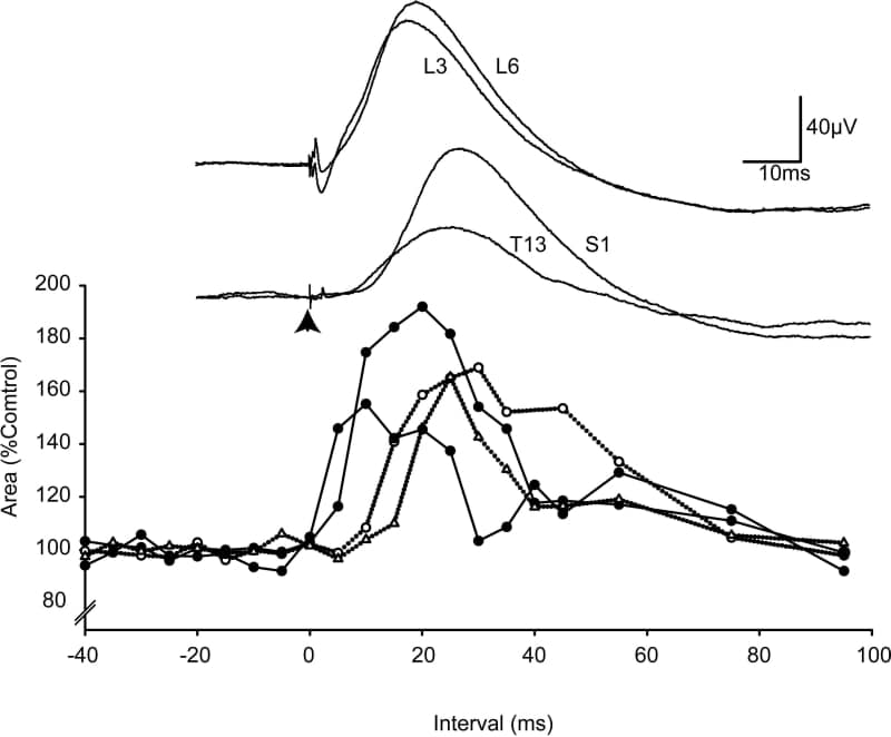
Fig 1. DRPs (above) and changes in sural nerve terminal excitability in response to stimulation of nearby and distant dorsal roots.
Where applicable, experiments conform with Society ethical requirements.