
In contrast to most Class III antiarrhythmic drugs that exhibit proarrhythmic side effects, amiodarone has been found effective and relatively safe. However, the electrophysiological mechanisms underlying the low arrhythmogenecity of amiodarone are poorly understood. This study compares electrophysiological properties of virtual ventricular tissues treated with amiodarone and d-sotalol, a Class III drug known to be proarrhythmic. A one-dimensional virtual ventricular wall is constructed based on the Luo-Rudy family of models for endocardial, M- and epicardial cells (Gima & Rudy, 2002). The effects of amiodarone (Sicouri et al. 1997) and d-sotalol (Akar et al. 2002) are incorporated as changes in the maximal conductances for the fast component of the potassium current IKr (the primary target for the Class III drug action) and the calcium current ICa,L, which reproduces the action potential duration (APD) changes in the three cell types. Results of computer simulations with the virtual wall are summarized in Fig. 1. d-Sotalol increases the transmural dispersion of repolarization in the wall by predominant increase of APD in M-cells, as seen by Akar et al. (2002). This widens the vulnerable window (VW) in the endo- and epicardial regions, where uni-directional block persists until the M-cell region is fully repolarized. Amiodarone, however, decreases the dispersion of repolarization, as seen by Sicouri et al. (1997), by prolonging APD in endo- and epicardial cells and decreasing APD in M-cells, which results in a narrow VW similar to that of a normal tissue. We conclude that the electrophysiological explanation for the safety of amiodarone in comparison to other Class III drugs lies in relatively low dispersion of repolarization leading to narrow VW, and hence, low probability of uni-directional block and initiation of re-entry in the ventricular wall.
University of Oxford (2004) J Physiol 561P, PC23
Communications: EFFECTS OF CLASS III ANTIARRHYTHMIC DRUGS ON VULNERABLE PROPERTIES OF VIRTUAL VENTRICULAR TISSUE
Aslanidi,Oleg V; Srinivasan,Neil T; Holden,Arun V;
1. School of Biomedical Sciences, University of Leeds, Leeds, United Kingdom. 2. School of Medicine, University of Leeds, Leeds, United Kingdom.
View other abstracts by:
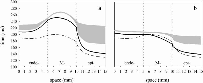
Figure 1. Effects of d-sotalol (a) and aniodarone (b) on the virtual ventricular wall. Spatial distributions of APD through the normal wall (dashed line) and the wall treated by drugs (solid lines) are shown along with the respective VWs (gray areas). Drug effects are modelled by differential blocking of IKr and ICa L in endo- M- and epicardial cells: for the d-sotalol model IKr is depressed by 40% in endo- by 100% in M-cell and by 65% in the epicardial region; for the amiodarone model IKr is uniformly depressed by 50% throughout the wall and ICa L is depressed by 40% in the M-cell region.
Where applicable, experiments conform with Society ethical requirements.