
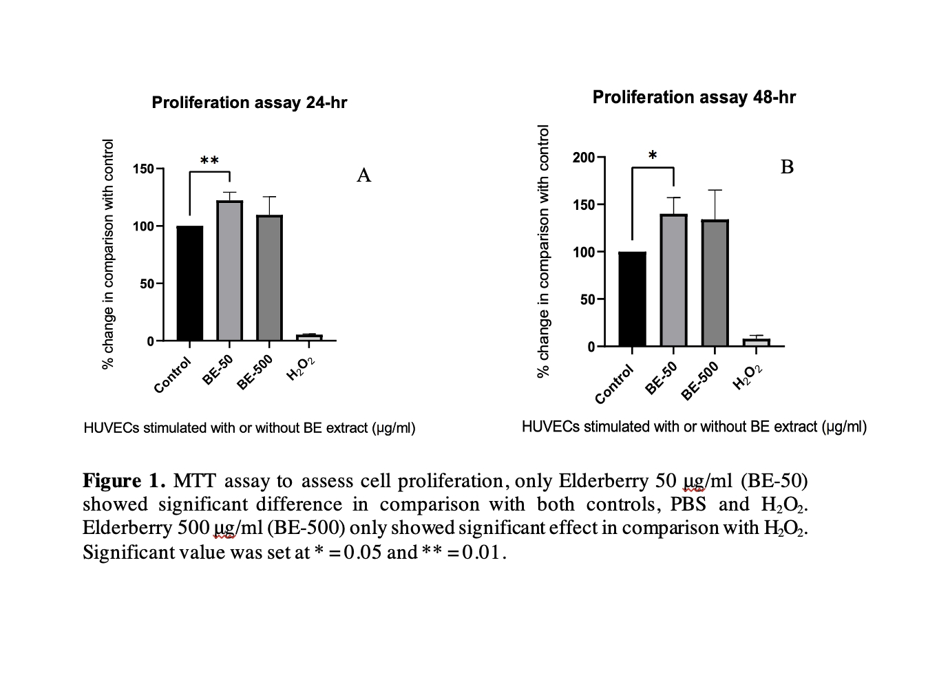
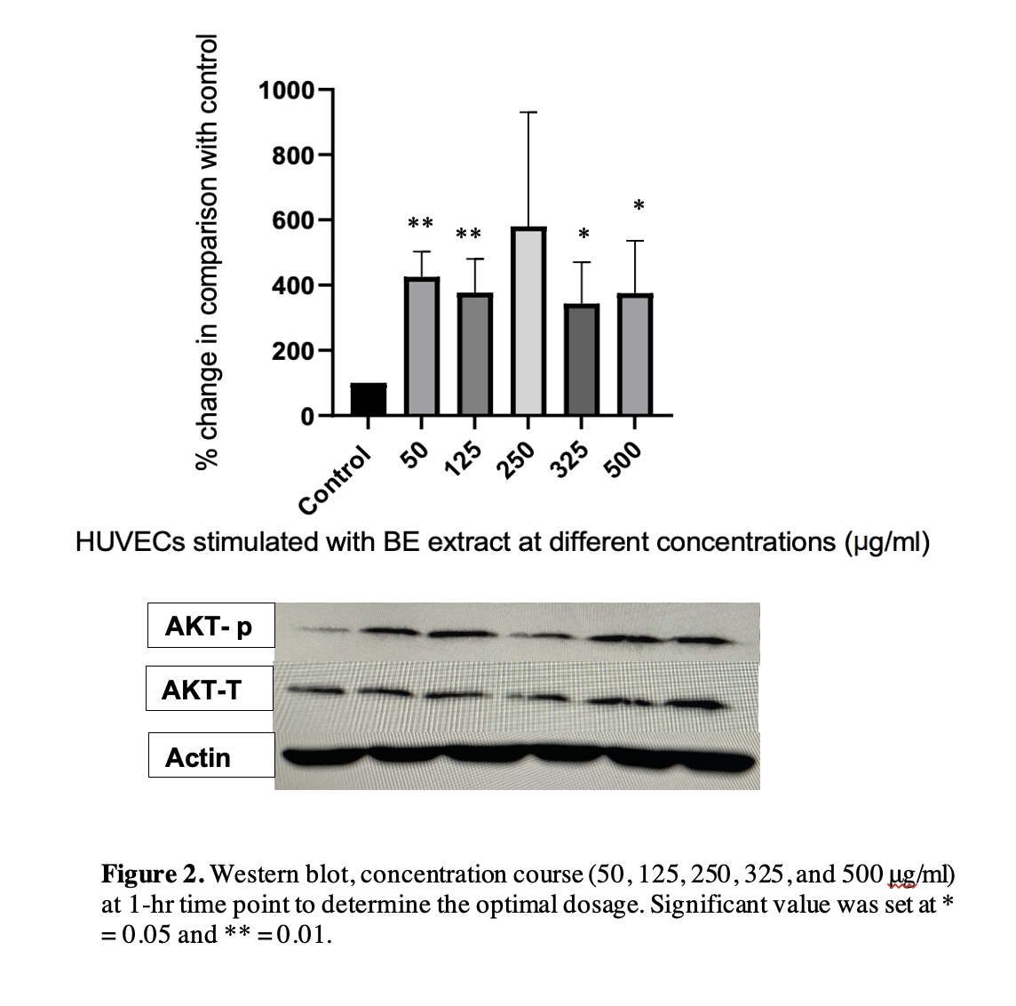
Introduction Previous research has shown that Elderberry extract (EE) can improve endothelial cell viability, which is vital for preventing endothelial dysfunction a subclinical marker for most cardiovascular diseases (1,2). However, the exact mechanisms have not been explored making it difficult for understanding application in vivo. Some research has postulated that AKT is related to cell survival (3). Objective Therefore, the main aim of this study was to confirm whether EE can improve endothelial cell viability and to explore the molecular mechanisms. Methods Primary Human Umbilical Vein Endothelial cells (HUVEC) were maintained in Media 200 supplemented with 10% foetal bovine serum and low serum growth supplement. The EE was dissolved in PBS. For the MTT assay cells were seeded at 3.5 x10³ per well in a 96 well plate, two concentrations were used including a low 50 μg/ml and high 500 μg/ml with two controls PBS and hydrogen peroxide (H2O2). The absorbance of each well was measured/quantified at 560 nM using a Promega GloMax Discovery system. For western blots, cells were treated with different concentrations (50, 125, 250, 325, and 500 μg/ml) for 1-hour and the protein levels of Phospho-AKT and AKT were measured by probing with specific antibodies. Levels of proteins were quantified and normalised using β-actin as an internal control, and the ratio of phosphorylated (activated) protein vs total protein was calculated. All data were analysed using GraphPad and were expressed as means ± standard deviation. Results H2O2 treated cells displayed a significant increase in cell cytotoxicity compared the control cells (p 0.05). Western blots were analysed by chemiluminescence detection using Li-Cor imaging system. For western blots time points which showed significant difference 125 μg/ml (p = 0.0098), 325 μg/ml (p = 0.0288), 500 μg/ml (p = 0.0412) although 50μg/ml the lowest concentration showed the largest change (F (7.821, 4) = 425.3 vs 100, p = 0.0019). Additionally, no difference was observed between the lowest and higher concertation (p > 0.05). Findings The main findings of this study show that a lower concentration of EE can cause proliferation within HUVECs and we also observed the highest AKT activity in the lowest concentration which could suggest there is an association. Further experiments are required with an AKT inhibitor (wortmannin) to confirm the association with AKT (4). Moreover, the PI3K/Akt pathway within endothelial cells has been known to induce eNOS phosphorylation which has many cardioprotective properties (5). Further research is required to understand the full functional properties of EE has within endothelial cells.
Future Physiology 2021 (Virutal) (2021) Proc Physiol Soc 47, PC04
Poster Communications: Elderberry extract increases cell proliferation in endothelial cells potentially via Akt pathway
Joseph Festa1, Mariasole Da Boit1, Aamir Hussain1, 2, Harprit Singh1
1 De Montfort University, Leicester, United Kingdom 2 University of Leicester , Leicester, United Kingdom
View other abstracts by:
Where applicable, experiments conform with Society ethical requirements.