
Prenatally, skeletal muscle and liver must prepare for new postnatal roles demanding extra energy, like locomotion and gluconeogenesis (1). Mitochondria are the primary source of this energy via oxidative ATP production. Fetal cortisol and tri-iodothyronine (T3) levels rise naturally towards term and stimulate maturation of tissues essential for neonatal survival (1). Both hormones also regulate adult metabolism (2,3). This study investigated the effects of these hormones on mitochondrial density in fetal ovine liver and skeletal muscle. All procedures were carried out under the Animals (Scientific Procedures) Act 1986. Under general anaesthesia, 12 single Welsh Mountain fetuses were catheterised at 118 days of gestation (d; term ~145d) and were infused with either cortisol (n=6; 2-3mg/kg/day) or saline (n=6) between 125-130d before euthanasia at 130d (200mg/kg sodium pentobarbitone, ewe and fetus). Another 12 twin-bearing ewes underwent thyroidectomy (TX) of one fetus and sham operation of its control twin under anaesthesia at 102d before euthanasia at 127d (n=6 ewes) or 143d (n=6 ewes). Samples of biceps femoris muscle (BF) and liver were collected from all fetuses and snap frozen. Fetal plasma cortisol was measured by ELISA and T3 by radioimmunoassay. Citrate synthase (CS) activity, a marker of mitochondrial density (4), was measured spectrophotometrically. Gene expression of PGC1α involved in mitochondrial biogenesis was measured by qRT-PCR. Data were analysed by t-test. Cortisol infusion significantly increased fetal cortisol levels at 130d (8.4±1.7 vs 49.2±2.4ng/ml, P<0.0001). Liver CS activity was significantly higher in cortisol than saline infused fetuses (Fig A), with a trend for higher PGC1α gene expression (P=0.07). However, at 130d, CS activity of the BF was unaffected by cortisol-infusion (Fig B). TX significantly reduced T3 levels at both ages (127d: 0.33±0.01 vs 0.12±0.03ng/ml; 143d: 0.60±0.14 vs 0.24±0.01ng/ml, P<0.05). At 127d, CS activity of the BF was significantly lower in TX than control fetuses, whereas TX had no significant effect on liver CS activity (Fig C & D). At 143d, CS activity was significantly lower in TX than control fetuses in both liver (P<0.05) and BF (P<0.001). Liver and skeletal muscle PGC1α expression was unaffected by TX at both ages. The results show cortisol and thyroid hormones have tissue specific effects on mitochondrial density in fetal ovine tissues at 127-130d, with cortisol more effective in liver than skeletal muscle. The reverse was seen with the thyroid hormones, although, by term, TX affected mitochondrial density in both tissues. Overall, the study suggests that both hormones are involved in prepartum preparation of mitochondria for the increased neonatal energy demands.
Physiology 2019 (Aberdeen, UK) (2019) Proc Physiol Soc 43, C084
Oral Communications: Endocrine regulation of fetal mitochondrial density in skeletal muscle and liver in fetal sheep
K. Davies1, R. M. Hess1, E. J. Camm1, A. J. Forhead1,2, A. J. Murray1, A. L. Fowden1
1. Department of Physiology, Development and Neuroscience, University of Cambridge, Cambridge, United Kingdom. 2. Department of Biological and Medical Sciences, Oxford Brookes University, Oxford, United Kingdom.
View other abstracts by:
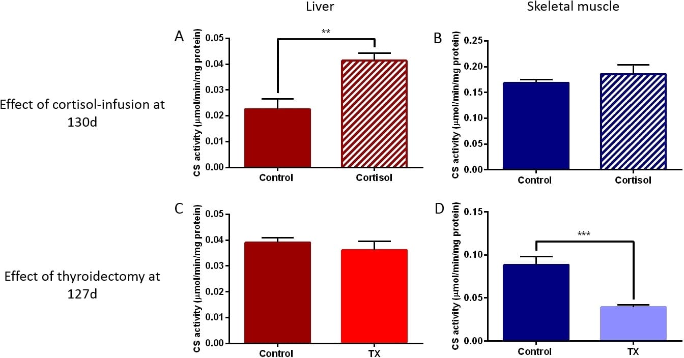
Figure. Mean�SEM citrate synthase (CS) activity in skeletal muscle and liver of A&B) control vs cortisol-infused fetuses at 130 days of gestation (d) and C&D) control vs thyroidectomised (TX) fetuses at 127d. ** P<0.01; *** P<0.001 by t-test.
Where applicable, experiments conform with Society ethical requirements.