
Introduction. Lactotrophs, neuroendocrine cells of the anterior pituitary, release peptide hormone prolactin from secretory vesicles through a fusion pore that is formed upon the fusion of the vesicle membrane with the plasma membrane. Spontaneous hormone discharge from a single vesicle is slower than under stimulation likely due to the kinetic constrains of the fusion pore openings (1). In the present work, we tested whether slow hormone release at rest could also be a consequence of a relatively narrow fusion pore. Methods. We monitored the permeation of fluorescent FM 4-64 dye molecules (diameter = ~1 nm) (1,2) and HEPES molecules (diameter = ~0.5 nm) through spontaneously forming fusion pores of prolactin vesicles by time-lapse confocal microscopy in resting lactotrophs, expressing synaptopHluorin (spH), a pH sensitive green fluorescent protein (3). Results. Exocytosis of prolactin vesicles (n = 36) in 10 mM HEPES solution resulted in a rapid increase in spH fluorescence, indicating an efflux of protons from acidic vesicles. We distinguished two populations of spontaneous fusion events, type I (Fig. 1A) and type II events (Fig. 1B). Type I events were devoid of FM 4-64 loading (n = 19, 53%), on the contrary in type II events vesicles loaded the FM 4-64 (n = 17, 47%). In both types of events, following the peak in spH signal, the signal either exponentially decayed (transient events; n = 23, Fig. 1), implying reacidification of the vesicle lumen due to the resealing of the fusion pore, or it remained at an elevated level during the monitoring (persistent events; n = 13), indicating that the vesicle lumen remained in contact with the extracellular space. Conclusions. The absence of vesicle entry by FM 4-64 dye in type I events and the absence of time-course modulation of these events in the presence of increased concentration of HEPES (100 mM), suggest that FM 4-64 and HEPES molecules did not pass the fusion pore in type I events (pore diameter 50% of the exocytic vesicles undergo fusion yielding subnanometer fusion pore diameters, precluding discharge of the hormone prolactin (diameter = ~5.2 nm).
Life Sciences 2007 (2007) Proc Life Sciences, C22
Research Symposium: Fusion pore opens to subnanometer diameters in resting neuroendocrine cells
N. Vardjan1, 2, M. Stenovec1, 2, M. Kreft2, 1, R. Zorec2, 1
1. Celica Biomedical Center, Ljubljana, Slovenia. 2. LN-MCP, Institute of Pathophysiology, Medical Faculty, University of Ljubljana, Ljubljana, Slovenia.
View other abstracts by:
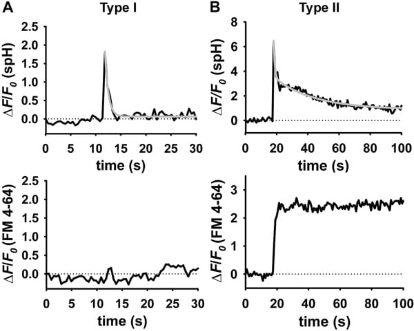
Fig. 1. Spontaneous exocytotic events of type I (A) and type II (B). Time-dependent fluorescence changes at the vesicle site separately for spH and FM 4-64. In type II events (B) FM 4-64 fluorescence simultaneously increased with the spH fluorescence signal.
Where applicable, experiments conform with Society ethical requirements.