
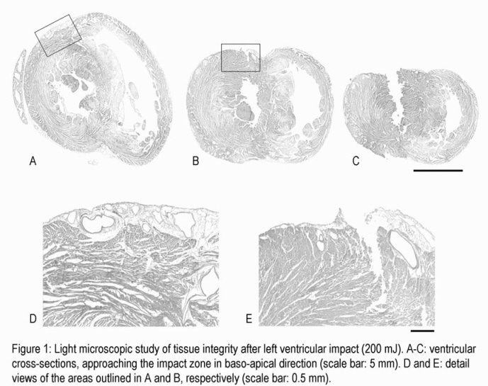
Investigations into the mechanisms of commotio cordis (mechanically induced dysrhythmia in the absence of structural damage [1]), are currently hampered by the lack of suitable isolated heart models. We have developed a mechanical stimulator for such studies (see demonstration at this meeting, [2]), and here we assess ventricular tissue integrity after mechanical interventions on humanely killed subjects, using histological techniques.Two types of mechanical impact were imposed: A) impact by a metal sphere after [3], and B) controlled mechanical stimulation / probe retraction as in [2]. After 1-3 impacts, ventricles were fixed by coronary perfusion with Karnovsky’s fixative (2% paraformaldehyde, 2% glutaraldehyde; pH 7.4). Impact sites were dissected out (with the exception of two hearts, where whole ventricular cross sections were obtained, Fig. 1) and embedded in paraffin wax. Sections (10 µm) were cut perpendicular to the epicardial surface, and stained with Mayer’s Haemalum, followed by an aqueous Eosin counterstain, prior to light microscopic examination.Initial experiments with method ‘A’applied impacts of 75 and 200 mJ. In contrast to a prior report, this did not cause only superficial tissue damage (<1 mm depth, [3]), but severe trauma including ventricular rupture (Fig. 1). Subsequent studies used method ‘B’to apply controlled impacts of 1-45 mJ. At energies above 20 mJ, clear compressive damage was identified in the tissue below the contact site, as well as (probably strain-induced) disruption surrounding that area. Light microscopic identification of impact-induced damage at lower energy levels, in particular near the threshold of 5-10 mJ (identified using creatine kinase [CK] assay [2]), proved difficult and could not always be concluded with certainty.In the given context, light microscopy would not appear to outperform CK assays for identification minor, mechanically induced tissue damage. Given the considerable difference in cost and time requirements, CK assays should form the ‘gold standard’ for routine assessment of tissue integrity in isolated heart studies of commotio cordis.
University of Glasgow (2004) J Physiol 557P, PC12
Communications: Histological validation of mechanically induced tissue damage in Guinea pig isolated heart
S.T. Schaaf (a),J.Sheldon (b), I.A. MacLeod (a),P.J.Cooper (a) and P. Kohl (a)
(a) University Lab of Physiology, Oxford, UK and (b) School of Biological and Molecular Sciences, Brookes University, Oxford, UK
View other abstracts by:
Where applicable, experiments conform with Society ethical requirements.