
We wish to study how visual motion-sensitive information is routed to and processed in Drosophila lobula plate tangential neurones (LPTNs) for understanding visual behaviour. Compared to higher organisms, the relatively simple and genetically malleable connections between Drosophila LPTNs, the eyes and brain can help us to make sense of motion coding strategies. Using transgenic flies which we have developed to be predominantly UV-sensitive while selectively expressing Ca2+-reporters (Cameleon) in their LPTNs (“UV-flies”, Figure 1), we have been able to record intracellular voltage and Ca2+ signals in LPTNs in response to moving light patterns in vivo. By genetically shifting the sensitivity of R1-R6 photoreceptors to UV-range, the visual excitation of photoreceptors does not overlap with the fluorescent genetic reporter, enabling us to undertake real-time Ca2+ imaging without influencing visual functions. Building on these efforts, we wish to now dissect the bottom-up and top-down connections to LPTNs, and their respective roles in computing neural representations of motion signals. This we plan to do by recording changes in voltage and Ca2+ signals of LPTNs to moving UV-stimuli while switching on/off different synaptic inputs to them. This can be achieved by using the UAS-Gal4 system to express light gated channels, NpHR to inhibit or ChR2 to excite neurons, or express temperature sensitive endocytosis blocking proteins, such as shibirets1. To undertake this task, we are constructing a hybrid instrument that combines 2-photon scanning microscopy and electrophysiology, capable of simultaneous measurements of voltage and Ca2+ signals in the tissue depths and spatial resolution that are required here. Furthermore, to correlate the changes in the network activity to the fly’s optomotor behaviour, our UV transgenic flies will be tested in a UV flight simulator system. The results and analysis of these experiments will be utilized for realistic mathematical modelling of motion detection.
University of Cambridge (2008) Proc Physiol Soc 11, PC3
Poster Communications: How does the Drosophila brain compute and see visual motion?
T. J. Wardill1, M. Juusola1
1. Department of Biomedical Science, University of Sheffield, Sheffield, United Kingdom.
View other abstracts by:
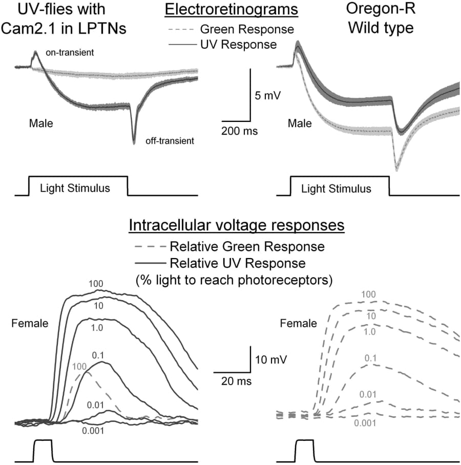
Figure 1. Responses of ‘UV flies’ (left) and wild type (right) to light pulses. The transients shown on electroretinograms (ERG) reflect synaptic transmission to large monopolar neurons (grey shading indicates S.E.). Wild type flies respond to both green and UV light due to the activation spectrum of Rhodopsin 1. ‘UV-flies’ expressing Rh4 in the place of Rh1 in photoreceptors R1-R6 show the greatest R1-R6 intracellular response to UV light with only small responses to very bright green pulses (light grey dashed traces). 'UV-flies' are >2 000 times more sensitive to UV than to green light (Wardill & Juusola unpub.).
Where applicable, experiments conform with Society ethical requirements.