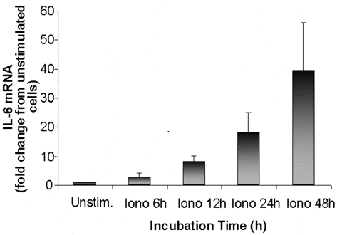
We have recently demonstrated that human skeletal muscle expresses and releases the cytokine interleukin (IL)-6 during exercise (Steensberg et al. 2001) and that the nuclear transcriptional activity of IL-6 is rapid after the onset of exercise (Keller et al. 2001). These data suggest that the contracting myofibres, rather than other cell types contained within skeletal muscle, are the source of muscle-derived IL-6 and, since calcium (Ca2+) is rapidly released from the sarcoplasmic reticulum at the onset of exercise, that the stimulus for IL-6 gene transcription may be via a Ca2+-dependent mechanism. To test these hypotheses, muscle biopsies were obtained from vastus lateralis of healthy young subjects. The study was approved by the local ethics committee in accordance with the Declaration of Helsinki. Biopsies were used to isolate satellite cells and grown into myotubes in culture. Muscle cell cultures were subsequently stimulated with the Ca2+ ionophore, ionomycin, for 6-48 h to result in an intracellular free Ca2+ level comparable to that during muscle contraction. IL-6 mRNA levels were determined by relative real-time PCR.
IL-6 (relative to β-actin, which was used as an internal control) increased progressively until the end of stimulation with a peak value of ~40-fold higher compared with that of unstimulated cells (see Fig. 1). These data demonstrate that myofibres are capable of producing IL-6 and that the rise in intracellular free Ca2+ is, at least in part, a mechanism responsible for the exercise-induced increase in IL-6 that we have previously observed.

