
Mechanisms underpinning ventricular fibrillation (VF) are poorly understood. Maintenance of VF by a single re-entrant source (mother rotor) or by multiple wavelets have both been implicated. We aimed to characterise the spatiotemporal dynamics of early VF in humans using total epicardial electrical mapping. In 10 patients undergoing cardiac surgery, VF was induced by burst pacing, and a 20-40 s episode of fibrillatory activity was sampled at 1 kHz using an epicardial sock containing 256 unipolar contact electrodes connected to a UnEmap system. Dominant frequencies (DF) were computed for each electrode signal using the fast Fourier transform [1]. We also transformed voltage into phase using the Hilbert transform. Phase singularities (PS) [2] and activation wavefronts (WF) [3] were identified and quantified. Regression analysis showed a small but significant increase in the mean DF at a rate of 0.018±0.005 Hz/s (p<0.01) during early VF across all electrodes of all hearts. Moreover, large wavefronts with significant organisation were present for the majority of VF recordings. Each patient had a median number of PS between 4 and 9, and a median number of WF between 3 and 7, indicative that early VF is driven by a small number of electrical sources. Our recordings demonstrated multiple electrophysiological activation patterns. For example, in the fibrillating heart of one patient, we observed (i) persistent clockwise rotation of a single epicardial re-entrant wave; (ii) persistent anti-clockwise epicardial re-entry (Fig. 1, left); and (iii) epicardial breakthrough patterns. Moreover, these organised patterns were punctuated by: (iv) disorganised wave activity with many interacting re-entrant sources (Fig. 1, right). Throughout the entire VF episode, the electrophysiological activity continually switched between these organised and disorganised behaviours. This type of transitional VF activity was observed for all patients. We found that early human VF was typically sustained by a small number of re-entrant sources that were associated with large and coherent WF (mother rotors). However, at times we also observed a large number of small WF (multiple wavelets). Thus, our results suggest that multiple electrophysiological mechanisms sustain VF in the human heart.
University College London 2006 (2006) Proc Physiol Soc 3, C23
Oral Communications: Human ventricular fibrillation: mother rotor or multiple wavelets?
Ayman Mourad1, Martyn P Nash1, Richard H Clayton2, Chris P Bradley3, Peter M Sutton4, Martin Hayward4, David J Paterson3, Peter Taggart4
1. Bioengineering Institute, University of Auckland, Auckland, New Zealand. 2. Department of Computer Science, University of Sheffield, Sheffield, United Kingdom. 3. Department of Physiology, Anatomy & Genetics, University of Oxford, Oxford, United Kingdom. 4. Departments of Cardiology and Cardiothoracic Surgery, University College London Hospitals, London, United Kingdom.
View other abstracts by:
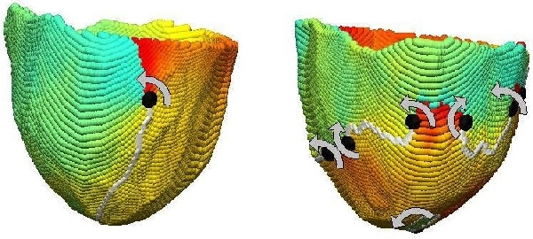
Figure 1. A single epicardial re-entrant wave (left) and multiple epicardial wavefronts (right) in the same patient. The spectrum shows electrophysiological phase and filled circles show phase singularities (with chirality indicated by the arrows) at the ends of the wavefronts (grey lines).
Where applicable, experiments conform with Society ethical requirements.