
During cell cycle progression, cells increase in size and must pass a size-dependent checkpoint prior to mitosis. Once a cell has passed this restriction point, it is committed to one cycle of division. Swelling could act as a proliferative signal through activation of cytoplasmic signalling cascades and gene transcription [1]. Mitogen activated protein kinases (MAPK) are central components of the signalling pathways involved in cell cycle progression. Previous studies from this laboratory have demonstrated a rapid early increase in ERK-1,2 activity in chick myocytes following hyposmotically-induced swelling [2]. Such MAPK activation could provide a link between swelling and proliferation. Embryonic chick hearts were removed at day 10 of development; myocytes were isolated enzymatically and maintained in culture. These cells may undergo at least one further cycle of cell division before terminal differentiation. Reducing extracellular osmolarity causes a significant increase in cell volume; e.g. cell volume increases to ~140% in hyposmotic solution (180 mosmol/l), before regulatory volume decrease occurs [2]. In this study, volume was perturbed by alteration of the extracellular osmolarity (150-400 mosmol/l by omission or addition of mannitol from the culture medium) for up to 48 hours. Cell proliferation was monitored by spectrophotometric assay of the number of viable cells [3]. Statistical comparison of absorbance between experimental groups was performed using one-way ANOVA and post-hoc Tukey tests; significant differences were accepted if p<0.05. Our results demonstrate that cells exposed to hyposmotic media (200mosmol/l) showed a rapid early increase in proliferation, compared to control cultures (Fig. 1). The absorbance was significantly increased, relative to controls at 24 h (0.172±0.071; n=9 wells cf. 0.001±0.004; n=6) and 48 h (0.300±0.095; n=12 cf. 0.048±0.033; n=8). In contrast, cells incubated in hyperosmotic media (400mosmol/l; n=2 experiments) failed to proliferate. Similarly, cells treated with the MEK inhibitor PD98059 (50μM) also failed to proliferate in hyposmotic media (n=2 experiments). This study establishes a link between hyposmotically induced cell swelling and an increase in cell proliferation, and suggests a role for ERK1,2 signalling in swelling-induced mitosis. These data demonstrate that inducing changes in cell size may accelerate or halt progression through the cell cycle.
University College London 2006 (2006) Proc Physiol Soc 3, PC176
Poster Communications: Hyposmotic cell swelling promotes cell division
Johanna E Parker1, Tim JC Jacob1, Sarah K Hall1
1. School of Biosciences, Cardiff University, Cardiff, United Kingdom.
View other abstracts by:
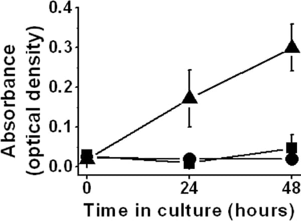
Figure 1. Cell swelling in hyposmotic media causes proliferation Circles culture medium (300mosmol/l); triangles hyposmotic media (200mosmol/l); squares isosmotic media (300mosmol/l; hyposmotic media + mannitol). Data shown as mean±sem of absorbance; n≥5 wells at each data point.
Where applicable, experiments conform with Society ethical requirements.