
Primary cilia are solitary, immotile, hair-like structures that protrude from the apical membrane of kidney epithelial cells into the cavity of the nephron. The function of the primary cilia was originally unknown, however recent work [1] has identified a link between primary cilia malfunction and autosomal dominant polycystic kidney disease (ADPKD). This malfunction suggests these organelles might act as flow sensors, signalling changes in luminal flow to control transepithelial ion transport along the nephron. Here we have adopted an interdisciplinary approach to investigate the structure and functionality of the primary cilium. We have imaged both Type I and Type II MDCK cells, using scanning electron microscopy (SEM) and atomic force microscopy (AFM). Cells were grown on glass coverslips and fixed with 2% glutaraldehyde in PBS. SEM was used to observe the density of cells and cilia, prior to AFM images being obtained of primary cilia on Type II MDCK cells. Live cells were imaged under liquid, and as the cilia remain mobile and not adherent to a substrate, their movement and orientation may affect the motion of the AFM cantilever. We found that it was possible to image unfixed cilia using AFM, by removing the cilia from the cell surface. We have achieved this by using a ‘peel-off’ method [2] whereby a poly-L-lysine-coated coverslip, placed on top of a layer of cells in a Petri dish, is removed in a single movement, and then imaged in situ. The nanoscale surface structure of the immobilised cilia can then be imaged with the AFM. From an analysis of AFM data, we have estimated cilium flexibility, and have probed cilium elasticity using AFM in a nanomechanical mode. By correlating AFM images with single-point AFM force curves, we observe a clear difference in stiffness between the cilium and the cell surface. Using a combined AFM/confocal microscope we have correlated the structures imaged by AFM, to fluorescent markers for tubulin and actin. The staining observed by confocal microscopy confirms that the structures protruding from the cells in the AFM images are the primary cilia. In a further development, we will report preliminary results from a combination of AFM imaging and patch-clamp electrophysiology to probe how cilium bending is coupled to ion transport.
University of Bristol (2008) Proc Physiol Soc 9, C4
Oral Communications: Imaging primary cilia on MDCK cells using atomic force microscopy
J. Evangelides1, D. N. Sheppard2, T. J. McMaster1
1. H. H. Wills Physics Laboratory, University of Bristol, Bristol, United Kingdom. 2. Department of Physiology and Pharmacology, University of Bristol, Bristol, United Kingdom.
View other abstracts by:
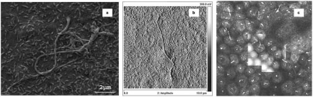
(a) SEM image (2 μm) of primary cilia on fixed MDCK cells; (b) AFM image (10 μm) of primary cilia on fixed MDCK cells; and (c) Combined confocal (immunofluorescence for tubulin and actin) and AFM images the latter present as an overlay in the centre of the confocal image.
Where applicable, experiments conform with Society ethical requirements.