
The spatial localization of Ryanodine receptor (RyR) clusters, and the number of RyRs per cluster affects the initiation and propagation of intracellular calcium waves [1]. Previous simulation studies have been based on simplified approximations of the distribution of RyR clusters. We embedded a three dimensional rat ventricular myocyte dataset (x,y: 0.05 µm; z:0.15 µm voxels) obtained by confocal microscopy [2] of part of an individual Z disk within a 300 by 450 by 40 cuboid of 0.05 µm voxels that extends into the adjacent sarcomeres, into a rat ventricular 3Dv E-Cell [3]. This represents part of a transverse slab, 2 µm thick, centered on the centre of the Z disk. To simulate spontaneous resting intracellular calcium dynamics we neglect voltage dependent sarcolemmal currents, and intracellular calcium dynamics were modelled by a set of reaction-diffusion equations based on Izu et al [1], with stochastic rules for triggering [Ca]2+ release and parameters given in [3]. The rate of change of [Ca]2+ depends on fluxes via RyR, pumps, leaks and buffers, where the firing probability of a RyR cluster is governed by probability function. In our simulations with diffusion coefficients of 7.9 µm2/s (transverse) and 17.1 µm2/s (longitudinal), sparse, localised regions/islands with high density of RyR clusters can act as foci for the initiation of propagating calcium events, and the peripheral region of the Z-disk produced more calcium release than the centre due to boundary effects.
University of Leeds (2008) Proc Physiol Soc 10, PC13
Poster Communications: Initiation and propagation of intracellular calcium waves within a three dimensional Z-disk of a rat ventricular virtual myocyte (3Dv E-cell)
P. Li1, C. Soeller2, M. Cannell2, A. V. Holden1
1. Institute of Membrane and Systems Biology, University of Leeds, Leeds, United Kingdom. 2. Department of Physiology and Anatomy, Faculty of Medical and Health Sciences, University of Auckland, Auckland, New Zealand.
View other abstracts by:
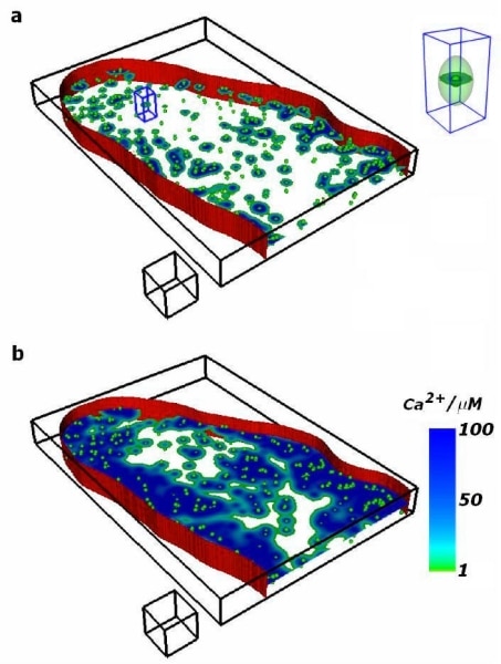
Figure 1 Transverse slab through part of the virtual cell encompassing the Z-disk the surface represents the sarcolemma and the balls demonstrate the spatial distribution of RyR clusters. (a) (t = 15 ms) and (b) (t= 65 ms) are frames from a movie illustrating initiation and propagation of intracellular calcium wave. The insert in (a) is a magnified view of the identified spark in (a) illustrating the anisotropy in the spread of a single spark. The spatial scale cube has the edge length of 2 µm.
Where applicable, experiments conform with Society ethical requirements.