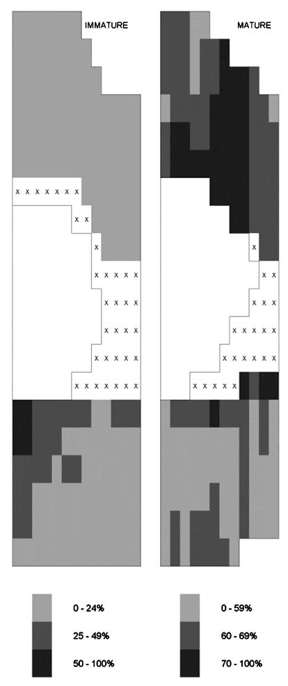
In immature rabbit aortas, atheromata develop preferentially in arrowhead-shaped regions immediately downstream of branch points. In mature rabbits, however, these regions are spared; disease occurs more frequently at the sides and upstream of the branches (Barnes & Weinberg, 1998). In immature rabbits, the permeability of the aortic wall to circulating macromolecules is greater downstream of branch points than upstream, and this pattern reverses with age (Sebkhi & Weinberg, 1996). The spatial correlation is consistent with permeability playing a critical role in atherogenesis. Although detailed maps of disease have been obtained, variation in permeability at the two ages has only been determined along the branch centre line. To test further the spatial correlation, we mapped permeability in a more extensive area around branch ostia.
All animal procedures complied with local and Home Office regulations. Male New Zealand White rabbits (Harlan IF strain, n = 4) were administered 700 mg kg-1 of rhodamine-labelled albumin I.V. heparin (2000 IU, I.V.) and an overdose (▓ge│ 500 mg, I.V.) of pentobarbitone was given 8 and 10 min later, respectively. The thoracic aorta was rapidly cannulated, flushed with saline and fixed in situ with formalin at physiological pressure. Regions surrounding intercostal artery branch ostia were embedded in epoxy resin and serial longitudinal sections 2 µm thick were cut from each branch, starting at its centre line. Tracer concentrations in the intima-media of every tenth section were measured by digital imaging fluorescence microscopy.
Tracer concentrations are shown for three branches from immature and for three branches from mature rabbits; maps for individual branches were normalized to give the same mean value before being combined.
These patterns of permeability are essentially identical to those we have previously reported for lesion frequencies in immature and mature rabbits, and to those reported for juvenile (Sinzinger et al. 1980) and young adult (Sloop et al. 1998) human aortas. The data strengthen the view that transport properties of the arterial wall play a key role in atherogenesis.
This study was supported by the BHF.
 View larger image [new window] |
| Figure 1. Each map shows tracer concentrations (% of maximum value in each map, X = not determined) in the aortic wall around half of an intercostal branch ostium (clear area). Mean aortic flow is from top to bottom. Length of each map is approx. 1 mm. |

