
The research investigates whether the location of mechanoreceptors contributes to the firing threshold using a continuum mechanics approach. Baroreceptors, situated in the media and adventitia of arterial walls, are considered. Histological studies have shown the media is elastically softer than the adventitia (J Levick, 2003). The main materials constituting these layers are elastin (elastically soft), collagen (elastically stiffer) and smooth muscle cells. Only the passive stress-strain relationship of elastin and collagen are considered in this study. Baroreceptors are classified as A- and C-fibres (Holzapfel et al., 2005). A-fibres are myelinated resulting in higher spike conduction speeds, whereas C-fibres are unmyelinated and slower. A-fibres have a conduction range in the region of 30-90 mmHg whilst type C has a range of 70-140 mmHg (J Levick, 2003). The relationship between blood pressure and strain energy, defined as the potential energy passively stored in a layer as a function of strain, in individual layers of a coronary arterial wall is explored. This is used to test the assumption that the threshold and sensitivity of the mechanoreceptor firing rate are affected by the stiffness of the tissue in which they are embedded. Mathematical models based on an empirically derived strain energy formulation were constructed for all three arterial layers, assuming a concentric cylindrical geometry. Each layer model incorporated factors such as heterogeneity, anisotropy, and collagen fibre angle. The same form was used for each layer, but with different material parameters. These were optimised using a Monte Carlo method using constraints defined by histological data and stress equilibrium (ME Mickael A Heydari, RS Crouch et al., 2010). The strain energy as a function of blood pressure for the adventitia and media of a coronary artery is shown in Figure 1. This shows that for a given blood pressure the adventitia stores more strain energy than the media (ME Mickael, A Heydari, S Pyner et al., 2010). Considering the case of baroreceptors with identical firing thresholds ( eg 1kJ) located in both these layers, the model predicts that the receptors in the adventitia would fire at a lower blood pressure ( e.g. ~30mmHg) than those in the media ( e.g. ~65mmHg). Similarly, if both receptors have the same saturation strain energy threshold, those in the adventitia saturate whereas those in the media have a much extended range. Thus the evidence suggests that receptors in the adventitia behave similarly to A-fibres whereas those in the media as C-fibres. Thus, this study concludes that it is theoretically possible for the location of a mechanoreceptor to affect the firing thresholds when expressed in terms of blood pressure.
Durham University (2010) Proc Physiol Soc 21, C04 and PC04
Oral Communications: Mechoreceptor firing threshold – A continuum mechanics study
M. E. Mickael1, A. Heydari2,1, R. S. Crouch1, S. Pyner2, S. Johnstone1
1. School of Engineering and Computing Sciences, Durham University, Durham, United Kingdom. 2. Biological and Biomedical Sciences, Durham University, Durham, United Kingdom.
View other abstracts by:
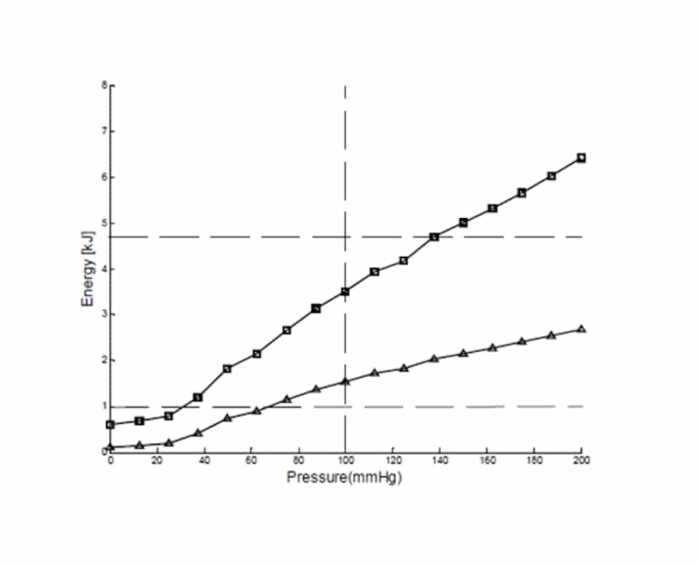
Figure1: Estimated strain-energy for the adventitia (squares) and media (triangle) as a function of luminal blood pressure for a coronary artery
Where applicable, experiments conform with Society ethical requirements.