
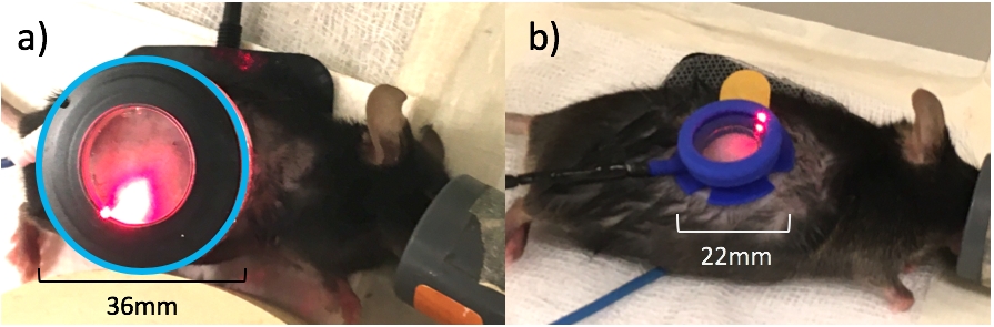
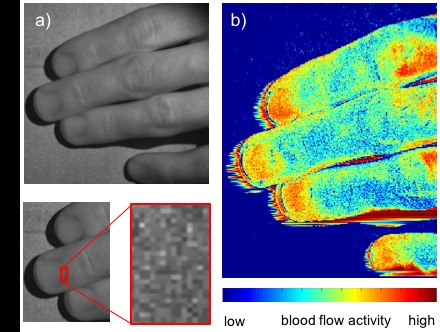
Objectives Endothelial dysfunction is important in the pathophysiology of preeclampsia often preceding the onset of the clinical disease, suggesting a major contributor to placenta dysfunction. Animal models offer distinctive opportunities to study both placental insufficiency and maternal endothelial function during pregnancy. New blood flow visualisation techniques are needed for phenotyping of murine models. We developed an endoscopic probe for real-time perfusion imaging of the placenta. We also miniaturized the hardware for transdermal drug delivery by iontophoresis to facilitate the assessment of maternal skin microvascular function in mouse models. Methods A minimally invasive probe featuring an endoscopic Laser Speckle Flow Imaging (LSFI) system to visualise real-time perfusion in organs was developed. LSFI is a real-time wide-field scanning technique, used clinically for assessment of skin microvascular function. Laser illumination of moving erythrocytes generates a dynamic pattern (speckles) based on backscattering properties, which quantify blood flow activity. For the application of iontophoresis in rodents we miniaturized the ion chamber, a device which is attached to the skin and contains the ionic drugs, houses the electric wiring, and allows optical blood flow imaging. The novel chamber has been designed, manufactured, and finally validated in a vasculature model of C57BI/6 mice. Results Our LSFI-device combines a miniature camera (speckle capture) with a fibre-coupled near-infrared laser (illumination) in the tip of the endoscopic probe. A micro camera (50%) and the use of a flexible material, it attaches better to small rodents. The weight reduction (by 80% to 2.5g total weight) is a significant refinement in pressure on the skin of small sedated animals. The smaller treatment area allows for faster and higher resolved LDI imaging and reduces the dose of delivered drugs. Hence, the systemic drug load on the animal is lowered and measurements are more cost effective. Conclusion The hardware for LSFI has been successfully miniaturized and tested in vitro. Use of the endoscopic laser to assess placental vascular insufficiency will be further validated in a mouse model of pregnancy. New iontophoresis hardware has been adapted and validated for the use in rodents. This technology will provide a platform to assess animal models of pregnancy and compare against clinical parameters to validate model characteristics. Endoscopic LSFI could provide wider clinical impact to visualize blood vessels and organ perfusion in real-time during surgery without the need for different imaging modalities or injection of dyes.
Physiology 2021 (2021) Proc Physiol Soc 48, PC071
Poster Communications: Microcirculation in a murine model of pregnancy: novel technique for endoscopic blood flow visualisation and optimised endothelial assessment
Lukas Markwalder1, Claire Sneddon1, Rodney Gush2, Nikola Krstajic1, Colin Murdoch1
1 University of Dundee, Dundee, United Kingdom 2 Moor instruments, Axminster, United Kingdom
View other abstracts by:
Where applicable, experiments conform with Society ethical requirements.