In the heart, cell swelling can occur during an acute period of ischaemia-reperfusion. Swelling has been shown to disrupt the microtubule (MT) cytoskeleton in embryonic cardiac myocytes (Larsen et al. 2000; Hall et al. 2001). Furthermore, MTs have been implicated in the regulatory volume decrease observed following swelling in embryonic myocytes (Hall et al. 2001). The aim of the present study was to investigate whether disruption of the MT cytoskeleton during hyposmotic challenge modulated cell swelling and contraction in the adult ventricular myocyte.
Hearts were removed from male Wistar rats following humane killing and ventricular myocytes were isolated by enzymatic disaggregation. Cells were incubated in either 10 mM colchicine (a MT disruptor) or the carrier solution methanol (0.1 %) for 2 h prior to functional studies. Control and colchicine-treated myocytes were stimulated at 0.5 Hz and superfused sequentially with normal physiological Hepes-based solution (290 mosmol), isosmotic solution containing sucrose and low Na+ (300 mosmol), hypo-osmotic solution (no sucrose, low Na+; 165 mosmol), and isosmotic solution (23 °C) (± colchicine). Changes in cell size were recorded from a video image of the cell, and cell shortening was measured by video-edge detection.
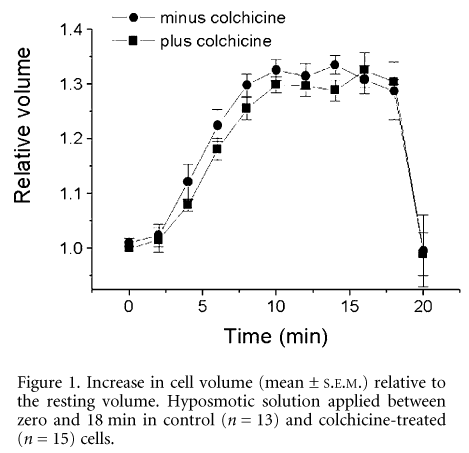
The mean maximal increase in cell volume of colchicine-treated cells and the time course of this effect, were not significantly different from control cells, during the 18 min exposure to the hyposmotic solution (Fig. 1). The contractile response of cells at maximal cell expansion, observed following 14 min exposure to hyposmotic solution, was not significantly altered for cells exposed to colchicine (P < 0.05, Student’s unpaired t test).
Microtubular disruption with colchicine did not significantly alter cell expansion in adult cardiac myocytes, in constrast with observations in embryonic myocytes (Hall et al. 2001). Data from this study suggest that colchicine-sensitive MTs do not modulate the volume or contractile response during hyposmotic swelling in the adult rat cardiac cell.
This study was funded by the British Heart Foundation (S.D.B. and S.C.C.).
All procedures accord with current UK legislation.

