The factors that affect the expression of genes coding for Ca2+ handling proteins in the heart are not well understood. We have, therefore, investigated the use of the muscle culture system described by Janssen et al. (1998) to study changes in mRNA levels in rat cardiac muscle.
Male Wistar rats (~300g) were killed humanely. A thin (< 1 mm) papillary muscle was dissected from the right ventricle and either frozen immediately in liquid N2, or after 8 h mounted in a muscle bath as described by Janssen et al. (1998), bathed in M199 culture medium containing 1.75 mM Ca2+ and equilibrated with 95 % O2-5 % CO2, stretched to Lmax and stimulated at 0.33 Hz using a stimulus ~10 % above threshold.
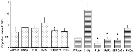
RNA was extracted using a Quiagen kit and reverse transcription carried out using Superscript II (Life Technologies) and random hexamers. Quantitative RT-PCR was performed using a Lightcycler (Roche) for the cDNAs of the following transcripts: 28S, sarcolemmal calcium ATPase (ATPase), calcineurin Aβ (CNAβ), phospholamban (PLB), ryanodine receptor (RyR2), sarcoplasmic reticulum ATPase (SERCA2a) and protein kinase Cα (PKCα). The amounts of mRNA were expressed relative to a standard sample and corrected for input with the value of 28S cDNA.Figure 1 shows that in freshly isolated muscle, the levels of the different mRNAs investigated were relatively uniform. However in the muscles maintained for 8 h, the levels of mRNAs coding for the Ca2+ handling proteins in the sarcoplasmic reticulum were significantly decreased.
These data suggest that this system provides a useful tool for study of gene expression in cardiac muscle, which will allow the effect of changes in environmental factors on gene expression to be investigated. The data also show that gene expression can change in 8 h following muscle isolation, which may have important implications for experiments on isolated tissue or cells maintained for 8 h or more. The factors causing the observed changes of expression are currently unknown, but the changes may represent reversion to a fetal pattern of gene expression, which is associated with the development of hypertrophy.
This work was supported by the British Heart Foundation.