
Multiphoton fluorescence microscopy is an exciting new optical sectioning technique which has great potential for numerous future developments and is ideal for applications that require deep optical sectioning of living tissue samples. In combination with microperfusion techniques, the major functions of the juxtaglomerular apparatus (JGA), the tubuloglomerular feedback (TGF) and renin release, can be studied with high spatial and temporal resolution. Salt-dependent changes in macula densa (MD) cell volume, vasoconstriction of the afferent arteriole (AA), and activity of an intraglomerular precapillary sphincter composed of renin granular cells are visualized in real-time. Imaging cytosolic calcium levels of the microperfused JGA dissected from kidneys from humanely killed rabbits, we observed a fast calcium wave using ratiometric real-time imaging with Fluo-4 and Fura-Red. This calcium wave initiated from the extra-glomerular mesangium and renin granular cells underneath the MD cells and was spreading towards both proximal AA smooth muscle cells and intraglomerular elements (mesangial cells and podocytes) with a time delay of 5 and 10 s, respectively. The terminal, intraglomerular part of the AA, a precapillary sphincter that includes renin granular cells, produced an almost complete closure of the AA during activation of TGF. This renin-positive sphincter acted as the first-response element of TGF activation and appeared to be the most significant vascular resistance to flow. In addition, release and tissue activity of renin can be studied on the individual granule level. Renin release from JG cells represents a unique form of exocytosis: even large granules can release their content very rapidly (within 300ms) and without any significant movement relative to the JG cell membrane. Using a novel, FRET-based fluorogenic renin substrate, we demonstrated interstitial renin activity around renin granular cells in response to the beta-mimetic isoproterenol, simultaneously with renin exocytosis. Imaging methods including the newest innovations in confocal fluorescence microscopy provide direct, visual information on JGA function with exceptional time and spatial resolution on the level of individual cells and organelles.
University of Bristol (2005) J Physiol 567P, SA43
Research Symposium: Multiphoton imaging of juxtaglomerular cell functions
Peti-Peterdi, Janos;
1. Physiology & Biophysics and Medicine, University of Southern California, Los Angeles, CA, USA.
View other abstracts by:
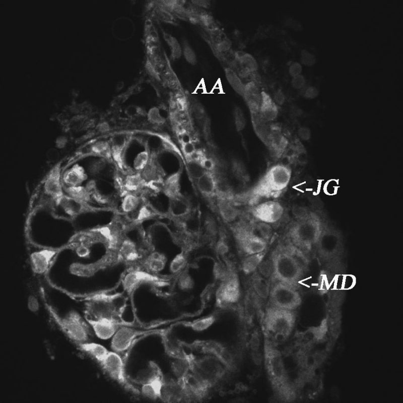
Figure 1. Two-photon image of a microperfused juxtaglomerular apparatus labelled with quinacrine. Diffuse labelling of the JGA structure including macula densa (MD) cells and the terminal part of the afferent arteriole (AA) containing renin granular cells or juxtaglomerular (JG) cells. JG cells contain a number of renin granules labelled with quinacrine.
Where applicable, experiments conform with Society ethical requirements.