Deep brain stimulation (DBS) is a common therapy for Parkinson’s disease. To determine clinically effective DBS parameters, clinicians complete extensive open-loop programming sessions which are prone to unreliability in accuracy. To guide parameter selection, neuroanatomical and neurophysiological biomarkers are being developed. At a neuroanatomical level, an increase in overlap between the volume of tissue activated (VTA) by the stimulation and target nuclei can be used to predict clinical outcomes [1]. Similar findings have been observed when using cortical recordings to guide DBS parameter selection [2]. Whilst both types of biomarkers can, to a degree, optimize DBS, they are not being used in conjunction as the relationships between them have not been fully explored.
This study aimed to determine the role of active electrode location, and corresponding overlap of the volume of tissue activated with the target nuclei, on cortical oscillatory activity during changes in DBS settings in Parkinson’s disease patients [3].
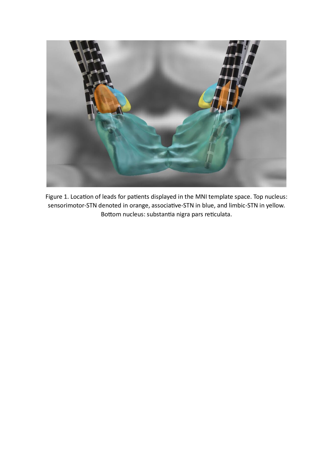
Using a 7-channel wireless EEG headset, cortical recordings were obtained during routine DBS parameter adjustment visits in the clinic (n=21). Arm tasks were used by the clinician to determine the efficacy of the DBS settings. From these task periods, relative power and bursting activity (burst amplitude, duration, and rate) were extracted for low (12-20Hz) and high (21-35Hz) beta power. To obtain the volume of overlap corresponding to the DBS settings, leads were localized by merging pre-operative MRI and post-operative CT scans in LEAD-DBS [4]. The location of the active electrodes was identified and overlap was calculated with the corresponding nucleus. If electrodes were in the subthalamic nucleus (STN) and an adjacent nucleus, overlap was calculated only with the STN. Following the extraction of these biomarkers, the delta between them for each pair of DBS settings was calculated.
Active contacts were found in the motor area of the STN (motor-STN; n=23), in the STN and adjacent nucleus (STN+; n=43), and in the substantia nigra pars reticulata (SNr; n=12). When stimulating multiple nuclei (STN+), change in low beta burst durations was reduced (p>0.01) compared to motor-STN stimulation. The low beta burst rate decreased when there was an increase in overlap with the motor-STN (p>0.01). High beta burst durations were greater when SNr was stimulated rather than motor-STN (p=0.03). High beta burst rates were significantly greater when active contacts were in the SNr than the motor-STN (p>0.01) or STN+ (p=0.02).
Neurophysiological biomarkers change depending on the location of the active electrode and which nucleus is being primarily stimulated. Low beta burst rate appeared most sensitive towards changes in overlap with the motor-STN but further investigation is required into other neurophysiological biomarkers.