
Aerobic system evaluation has many clinical applications. Commonly, the aerobic system is investigated by the analysis of muscular oxygen uptake (VO2m) dynamics through the modeling of the alveolar oxygen uptake (VO2a) responses during exercise. However, VO2m dynamics differ from VO2a due to circulatory distortions. Therefore, new methods are needed to estimate VO2m during exercise. The goal of this study was to develop a method for VO2m estimation based on near-infrared spectroscopy (NIRS, VO2m-NIRS). The VO2m-NIRS dynamics were then compared with VO2a and VO2m dynamics estimated by the circulatory method (VO2m-CM) [1]. Twenty-three participants (12 men and 11 women, 27±4 years old, 69±11 kg and 170±9 cm) performed a pseudo-random binary sequence exercise protocol (PRBS). Changes in concentrations of the vastus lateralis oxy- and deoxy-hemoglobin (O2Hb and HHb, respectively) were measured by the Oxymon device (ARTINIS, The Netherlands). The VO2a was measured by a metabolic cart while VO2m-CM was estimated following a previously published circulatory model [1]. The VO2m-NIRS was estimated from the O2Hb/tHb signals (Figure 1A), where tHb=O2Hb+HHb. The NIRS data were filtered and detrended, and then a peak detection algorithm was used to identify each contraction/relaxation phase. The slope of the O2Hb/tHb data for each contraction was computed (m in Figure 1B) and interpreted as VO2m since it calculates the O2Hb decay rate normalized by the local blood volume (i.e., tHb). The VO2a, VO2m-CM and VO2m-NIRS dynamics were analyzed by a cross-correlation function (CCF in Figure 1C) between the time-series of these variables and the PRBS workload (“Input” in Figures 1A and 1B). The CCF peak (CCFpeak, or the response speed) and lag (CCFlag, or the response delay), as aerobic system evaluation indexes, were compared between VO2a, VO2m-CM and VO2m-NIRS by repeated measures ANOVA. The correlation level was calculated by Pearson’s correlation coefficient. The statistical significance level (p) was set at 0.05 (two-tailed). As expected from a local VO2m measure, the CCFlag of VO2m-NIRS occurred statistically (p<0.05) before VO2m-CM and VO2a. In addition, the VO2m-NIRS CCFpeak was statistically (p<0.05) correlated with the CCFpeak of VO2m-CM and VO2a (Figure 1 D and E, respectively). The NIRS-derived VO2m (i.e., VO2m-NIRS) seemed to be reliable for the investigation of VO2m dynamics during exercise, therefore, the aerobic system can be evaluated directly from NIRS data.
Physiology 2019 (Aberdeen, UK) (2019) Proc Physiol Soc 43, PC092
Poster Communications: NIRS-derived Muscular Oxygen Uptake During Exercise in Humans
T. Beltrame1,2,3,4, J. Koschate2, U. Hoffmann2, M. Gois3, R. Hughson5, M. Frade3, S. Linares3, R. Torres1, A. Catai3
1. University of Campinas, Campinas, Brazil. 2. German Sport University Cologne, Köln, Germany. 3. Federal University of Sao Carlos, Sao Carlos, Brazil. 4. Universidade Ibirapuera, So Paulo, Brazil. 5. University of Waterloo, Waterloo, Ontario, Canada.
View other abstracts by:
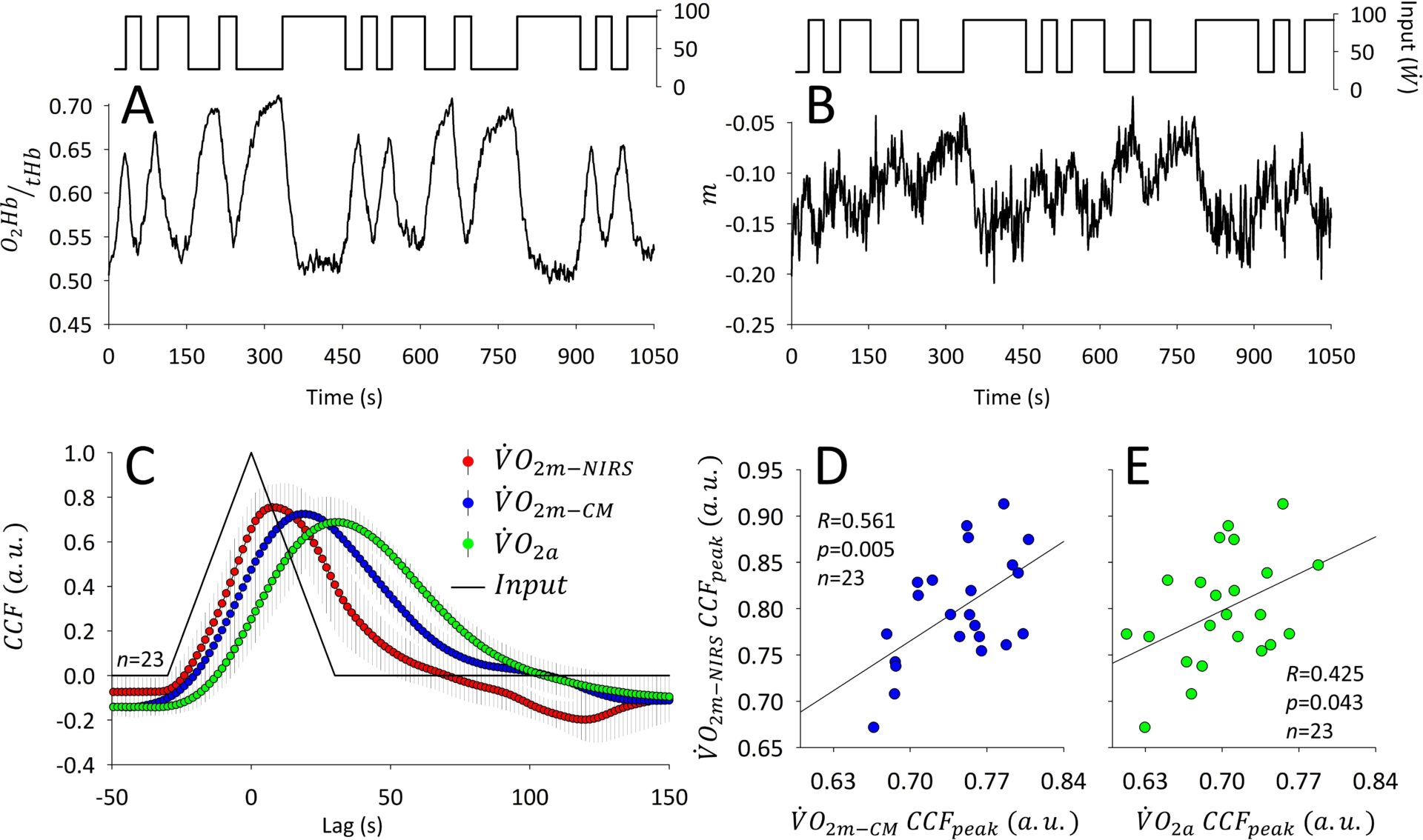
Method for muscular oxygen uptake estimation from near-infrared spectroscopy during exercise. Please see text for further details.
Where applicable, experiments conform with Society ethical requirements.