It is known that myocardial fibres rotate counterclockwise through the wall of the heart. Abrupt rotations in fibre orientation are implicated in arrhythmogenesis and should be considered during measurements of conduction velocity in ventricular myocardium. We combined histology, optical mapping and computer modelling to test the hypothesis that abrupt changes in subepicardial fibre orientation are responsible for unusual diamond-shaped activation patterns in swine RV.
Young pigs (n = 7) were heparinized (500 IU, I.V.) and subsequently anaesthetized with sodium pentobarbital (35 mg kg-1 I.V.). All procedures were conducted in agreement with the Guide for the Care and Use of Laboratory Animals (NIH publication no. 85-23, revised 1996). The heart was quickly removed and the RV free wall excised and cannulated through the right coronary artery. Preparations were then perfused and superfused with an oxygenated Tyrode solution, and stained with the voltage-sensitive dye di-4-ANEPPS. Contractions were inhibited using diacetyl monoxime (15 mmol l-1). Optical signals were recorded from the epicardial surface with a CCD video camera at 800 fr s-1. After completion of the experiments preparations were sectioned using traditional histological methods. Fibre orientation of each section was determined using an automatic algorithm. To simulate voltage-dependent optical signals, measured fibre angles were introduced into a computer model comprised of a three-dimensional model of electrical propagation and a multi-scattering light transport model. Electrical action potential waveforms were generated using Fenton-Karma kinetics. Optical action potential waveforms were obtained by convolving optical point spread functions, derived from the light transport model, with the computed three-dimensional distribution of the transmembrane potential.
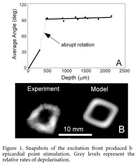
In the majority (n = 4/7) of preparations, we observed an abrupt change in fibre angle (70-90 ± 10 deg (S.D.)) at subepicardial depths of ~500 mm (Fig. 1A). These large magnitudes of rotation produced unusual diamond and quasi-rectangular-shaped activation patterns during epicardial point stimulation. The shape of wavefront directly correlated with the amount of rotation observed, with diamond shapes displaying the most rotation. Insertion of histological data into computer models accurately reproduced experimental observations (Fig. 1B).
Fibre rotation in swine RV is non-uniform, resulting in unusual patterns of electrical activation on the epicardial surface. This is important for understanding electrical propagation in the three-dimensional wall of the heart, and fibre architecture in the RV free wall.
This work was supported by Grant 2PO1-HL-39707 from the National Heart, Lung and Blood Institute of the National Insitutes of Health.
All procedures accord with current National guidelines.