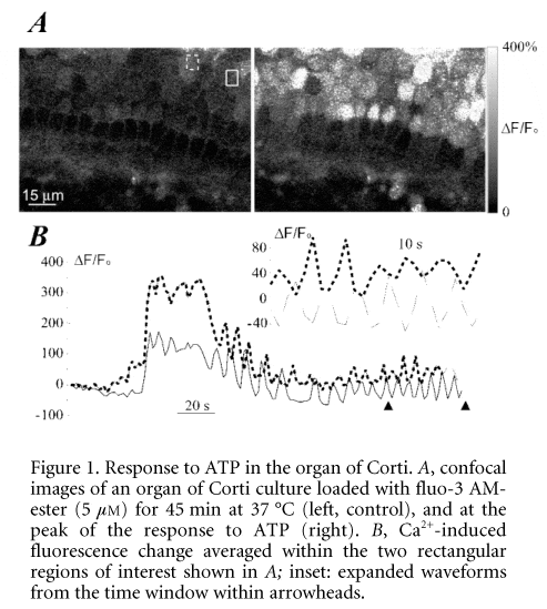
The level of ATP in cochlear fluid has been shown to increase in response to sound over-stimulation. The two most prominent types of supporting cells in the organ of Corti, Deiters’ and Hensen’s cells, have been shown to respond to focal application of ATP with transient elevation of intracellular Ca2+ levels (Lagostena et al. 2001; Lagostena & Mammano, 2001). Here we used an organotypic preparation from humanely killed rats (Sobkowicz et al. 1975) of the immature cochlea (prepared on postnatal day 1 and utilised up to 2 days in culture) to follow the intercellular spread of ATP-induced Ca2+ signals. Bolus application of ATP (final concentration 50-100 mM) in Ca2+-free HBSS (containing (mM): KCl 5.33; KH2PO4 0.44; NaHCO3 4; NaCl 138; Na2HPO4 0.3; glucose 5, 6; pH 7.4) produced an initial generalised increase of intracellular Ca2+ concentration (Fig. 1A), followed by generation of oscillatory Ca2+ responses in selected supporting cells (Fig. 1B).
In a given cell, oscillations occurred with a frequency of 0.163 ± 0.012 Hz (n = 4, mean ± S.E.M.). A clear pattern emerged during the oscillatory phase of the responses, characterised by intercellular propagation of Ca2+ waves. Propagation velocity was 9.6 ± 1.4 mm s-1 (n = 18). Oscillatory responses were either absent or greatly attenuated if Ca2+ (2 mM) was included in the extracellular medium. Facilitation of ATP responses in Ca2+-free medium is consistent with waves being carried by a mechanism that involves ATP-induced ATP release through open connexin hemichannels (Cotrina et al. 1998).
This work was supported by grants from the INFM (R.L. TSB2) and MIUR to F.M. and Tullio Pozzan. V.P. and C.D.C. were supported by fellowships from the IRCSS S.G.R. and UNIDO, respectively.
All procedures accord with current National and local guidelines.