
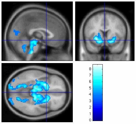
Neurodegeneration with brain iron accumulation 1 (NBIA 1: formerly known as Hallervorden-Spatz disease) comprises a rare group of autosomal recessive extrapyramidal movement disorders accompanied by iron accumulation in the globus pallidus and nigrostriatal pathways and mutations in the pantothenate kinase (PANK2) gene [1,2]. Here, we demonstrate the cerebral functional metabolic correlates in patients with NBIA1, assessed prior to deep-brain stimulation (DBS) [2]. Brain glucose metabolism was measured using 18F-fluoro-deoxyglucose positron emission tomography (FDG-PET) using high-sensitivity 3D acquisition in five children aged 4, 9, 10, 13 & 14 years old with late infantile PANK2 disease. Regional differences in brain glucose uptake were analysed by statistical parametric mapping using SPM2 software (www.fil.ion.ac.uk) to compare subject scans with a normative database of 20 subjects aged 40-60 years. All the PANK2-positive PET-CT scans were considered normal on routine clinical inspection. Between groups SPM2 comparison showed significant cerebral hypometabolism within bilateral globus pallidus, cerebral peduncles, occipital cortex, cerebellar vermis, and pons (p < 0.001). The statistical parametric maps of hypometabolism for the group of five cases is shown superimposed on an average of 156 MRIs in MNI (Montreal Neurological Institute) space in sagittal (a), coronal (b) and axial (c) planes. Late infantile PANK2-positive NBIA1 is associated with decreased metabolism within bilateral visual cortex, globus pallidi, cerebral peduncles, dorsal pons and cerebellar vermis. Although further comparisons with age-matched paediatric controls are required, hypoperfusion of the head of the right caudate nucleus, pons, cerebellar vermis have been previously reported in one case with PANK2 disease [3]. Hypometabolism extending beyond the pallidal-substantia-nigra abnormalities identified on MRI could reflect functional or structural deficits. If functional, they may provide a measure of response to DBS.
University College London 2006 (2006) Proc Physiol Soc 3, PC59
Poster Communications: Patterns of abnormal cerebral metabolism in late-infantile PANK2 mutation-positive neurodegeneration with brain iron accumulation type 1 (NBIA 1) (Hallervorden Spatz Disease) measured using 18F-fluoro-deoxyglucose positron emission tomography and statistical parametric mapping
Jean-Pierre Lin1, Laurence J Reed2, Richard Selway3, Huma Sethi3, Michael Samuel3, Jozef Jarosz3, Gonzago Alarcon3, Joel Dunn4, Edward Somer4, Waja
1. Paediatric Neurology Complex Motor Disorders Research Group, Evelina Children's Hospital, Guy’s & St Thomas’ NHS Foundation Trust, London, United Kingdom. 2. Division of Psychological Medicine, Institute of Psychiatry, King’s College London School of Medicine, London, United Kingdom. 3. Clinical Neurosciences, King’s College London School of Medicine, London, United Kingdom. 4. The PET Imaging Centre at St Thomas’s Hospital, King’s College London School of Medicine, London, United Kingdom. 5. Clinical Radiology Evelina Children’s Hospital, Guy’s & St Thomas’ NHS Foundation Trust, London, United Kingdom.
View other abstracts by:
Where applicable, experiments conform with Society ethical requirements.