
The complex electrophysiological behaviour of the mammalian atrioventricular node (AVN) compared to other regions of the heart is well known. Numerous clinical investigations have described various electrophysiological phenomena unique to the human AVN. However, little is known about the molecular nature of ionic currents responsible for this electrical activity. The aim of our study was to use quantitative PCR (QPCR) to identify ion and gap junction channels and membrane protein subunits governing the electrical activity in the human AVN. Three AVN samples (with surrounding working myocardium) were cryosectioned and specific sub-regions: compact node (CN), His bundle (HB), atrial muscle (AM) and ventricular muscle (VM)] were microdissected. Total RNA was extracted from these regions and cDNA synthesised, and used for QPCR analysis (ABI 7900HT). Hearts were obtained from the Queensland Heart Valve Bank. The ion channel transcripts in Table 1 were analysed for the level of abundance across different tissue types, and amongst each group of transcripts within each tissue type. For example, when compared to different tissue types, Cx43 was the least abundant in the CN. However, when compared to different connexins within the CN, Cx40 was the most abundant. Interestingly, out of all transcripts investigated, the Ca2+ handling proteins showed the most abundant mRNA levels in the working myocardium as well as the AVN, whereas amongst the ion channels (after HERG), the twin-pore K+ (TWIK1), together with HCN4, appear to be the second most abundant ion channel subunits in the human AVN. We have shown that the two sub-regions of the human AVN to contain a unique ion channel expression profile compared to working myocardium. Our data provide insights into the electrical function of the AVN.
University of Manchester (2007) Proc Physiol Soc 8, PC27
Poster Communications: Prediction of the electrical activity of the human atrioventricular node using a quantitative PCR-based approach
I. D. Greener1, N. J. Chandler1, J. O. Tellez1, P. Molenaar2, D. C. Sigg3, V. Sharma3, M. R. Boyett1, H. Dobrzynski1
1. Cardiovascular and Endocrine Sciences, University of Manchester, Manchester, United Kingdom. 2. University of Queensland, Queensland, QLD, Australia. 3. Medtronic Inc., Minneapolis, MN, USA.
View other abstracts by:
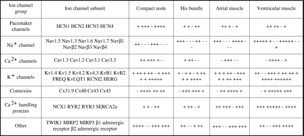
Table 1. Summary of expression profile of ion channel and gap junction channel, and Ca2+ handling protein subunits throughout cardiac tissues.
Where applicable, experiments conform with Society ethical requirements.