
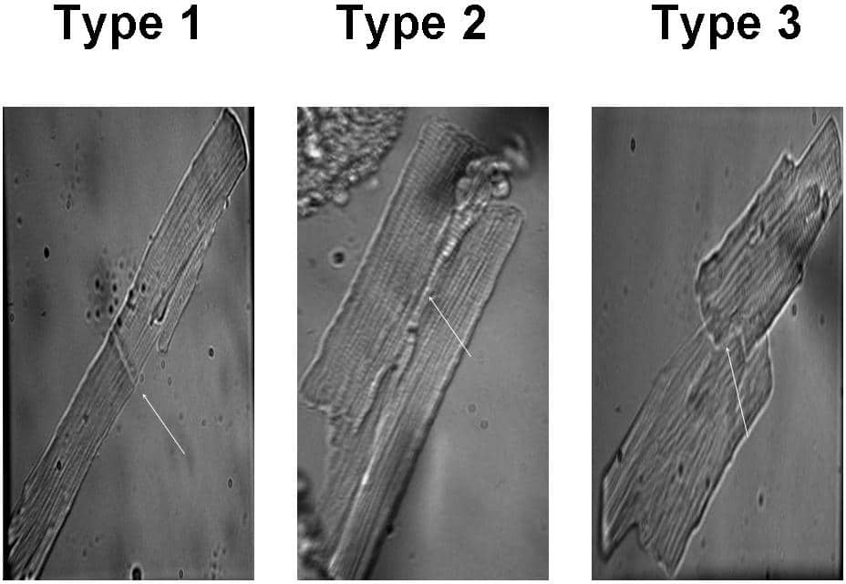
Systolic alternans presents in the final stages of heart failure and is thought to provide the arrythmogenic substrate that kills about 40% of the patients at this stage of the disease. Systolic alternans has been potentially linked to cardiac arrhythmias including ventricular tachycardia and ventricular fibrillation. We previously have shown that intracellular Ca wave propagation in a single myocyte is involved in alternation of the systolic Ca transient 1-3. These data have so far come from studies in single cells but in the whole heart there must be some way to provide for propagation between cells. However, there is little information about the intercellular propagation of Ca waves between cardiac myocytes. Therefore, the aim of this study was: 1) to observe the propagation of Ca waves between paired ventricular myocytes; 2) to investigate the mechanism involved in the propagation of alternans between myocytes. Fig.1. Three representative paired ventricular myocytes. Type 1 pairs are connected in an end-to-end fashion by relatively simple intercalated disks (white arrows) without complex staircase configuration characteristic of ventricular myocytes as in type 3 pairs. Three types of electrically coupled cells were studied. Type 1-3 as shown in Fig. 1. Propagation of spontaneous Ca waves between cells were observed in 7 out of 9 in type 2 pairs and 6 out 6 in type 3 pairs. Although spontaneous waves of Ca release did occur in Type 1 cell pairs propagation from one cell into the other never occurred. By applying a small depolarising pulse to one cell of the pair, subcellular alternans can be successfully produced in all three pair types; whereas a propagating Ca wave travelling from one cell to its neighbour was never seen in type1, it was seen in 5 out of 6 in type 2 and 6 out of 6 in type 3. Propagation of Ca waves between type 2 and type 3 pairs is generally synchronized. The lack of communication between type 1 paired cells is going to be further investigated by measuring conductance of gap junctions. Relatively lower expression of connexin in the intercalated disk may contribute to poor coupling between type 1 paired cells 4, in addition the distance to the nearest SR Ca release unit might be too far from the intercalated disc to support propagation.
University of Manchester (2010) Proc Physiol Soc 19, PC122
Poster Communications: Propagation of calcium waves between paired ventricular myocytes from rat
Y. Li1, D. A. Eisner1, S. C. O'Neill1
1. Cardiovascular Medicine, Manchester University, Manchester, United Kingdom.
View other abstracts by:
Where applicable, experiments conform with Society ethical requirements.