
Accurate simulation of the generation and propagation of cardiac electrical activity requires detailed electrophysiological and anatomical models. We have previously generated three-dimensional (3D) anatomical models of the sinoatrial node and the atrioventricular node (Dobrzynski et al., 2005; Li et al, 2004). The purpose of this study was to construct a 3D anatomical model of the rabbit right atrium with multiple objects, including the conduction system, and use this to simulate the propagation of the action potential through the right atrium. In this work, we used multiple techniques to generate the 3D anatomical model. The model was based on ~1500 images of the rabbit heart (atria and part of ventricles) obtained by high-resolution magnetic resonance imaging (MRI). MATLAB was used to analyse the data and generate a 3D anatomical model of the right atrium. The ~1500 two-dimensional images were stacked to produce a 3D array. Segmentation was carried out semi-manually by analysing the data using custom written programs developed in MATLAB. The sinoatrial node (SAN) and atrioventricular node (AVN) could be detected (and then segmented) after comparison of images with our existing models of the SAN and AVN. A 3D right atrium array model, including thirteen objects, was constructed. The objects are the right atrium, the SAN, the AVN, part of the right ventricle, the aorta with the aortic valve, the superior vena cava, the inferior vena cava, the coronary sinus, the tricuspid valve, part of the mitral valve, the fossa ovalis, the central fibrous body and outer fatty and connective tissue. The FitzHugh-Nagumo model was used to simulate the propagation of the action potential from the SAN through the 3D right atrium model. The propagation sequence is shown in Fig. 1.
University of Manchester (2007) Proc Physiol Soc 8, PC31
Poster Communications: Propagation of the action potential through a 3D model of the rabbit right atrium including the conduction system
J. Li1, J. E. Schneider2, I. D. Green1, H. Dobrzynski1, M. R. Boyett1
1. University of Manchester, Manchester, United Kingdom. 2. University of Oxford, Oxford, United Kingdom.
View other abstracts by:
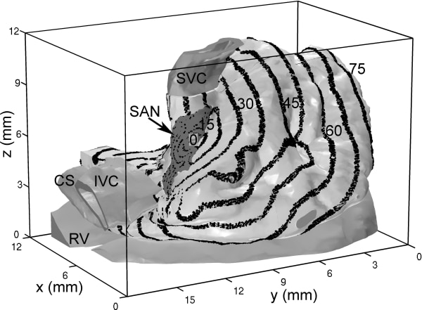
Figure 1. The propagation sequence of the action potential through the 3D right atrium. Isochrones are shown at 7.5 ms intervals.
Where applicable, experiments conform with Society ethical requirements.