
Clinical trials show that Class III antiarrhythmic drugs (e.g., d-sotalol) have proarrhythmic side effects, and only amiodarone is relatively safe. At the cellular level, the drugs have different effects on the transmural dispersion of action potential duration (APD), which d-sotalol increases and amiodarone decreases. Our previous studies [1] suggested that such differences can account for the safety of amiodarone at the tissue level, as smaller APD dispersion results in proportionally small vulnerable window (VW). In this study we quantify the effects of d-sotalol and amiodarone using detailed models, which are validated against respective experimental results for the canine ventricular tissue [2]. The Hund-Rudy [3] canine ventricular excitation model has been modified to reproduce the electrical restitution properties of endo-, M- and epicardial cells [4], as well as experimentally observed changes of these properties under the action of amiodarone and d-sotalol [2]. The resultant cell models were then incorporated into tissue models: a 1D strand model for transmural propagation, and a 3D wedge based on diffusion tensor MRI reconstruction of the canine ventricular architecture [5]. The standard S1-S2 protocol was used to measure vulnerability of the tissue defined as the time width of VW during which the S2 stimuli produced unidirectional conduction block. The models generate transmural APD dispersion patterns that are consistent with experiments on dogs treated with amiodarone and d-sotalol. The profile of VW is spatially heterogeneous, and correlates with the transmural APD dispersion (Fig. 1); VW is primarily wide in the epicardial region, where unidirectional block persists until the M-cells are fully repolarised. As d-sotalol increases and amiodarone decreases the APD dispersion, the VW in the latter region becomes proportionally larger or smaller (at maximum around 30 ms with d-sotalol, compared to 7 ms with amiodarone). S2 stimulation in the epicardial region of the 3D wedge within the VW results in sustained re-entry in case of d-sotalol; however, re-entry fails in control or with amiodarone. In conclusion, our results provide an electrophysiological explanation for the relative safety of amiodarone in comparison to d-sotalol, and quantify the vulnerability to re-entry in the canine ventricular tissues affected by the Class III drugs.
University College London 2006 (2006) Proc Physiol Soc 3, PC92
Poster Communications: Quantifying the effects of Class III drugs on the canine ventricular tissue
Oleg V Aslanidi1, Alan P Benson2, Arun V Holden2, Henggui Zhang1
1. School of Physics and Astronomy, University of Manchester, Manchester, United Kingdom. 2. Institute of Membrane and Systems Biology, University of Leeds, Leeds, United Kingdom.
View other abstracts by:
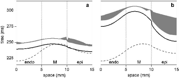
Figure 1. Transmural dispersion of APD90 (solid line) and spatio-temporal extent of the VW (grey area) in the 1D canine ventricular strand: (a) amiodarone and (b) d-sotalol; dispersion of APD90 in control is shown by dashed lines. The effect of d-sotalol is modelled by depressing IKr by 50% in endo- by 70% in M- and by 20% in the epicardial cells; the effect of amiodarone is modelled by depressing IKs by 80% in endo- and by 30% in epicardial cells and depressing INa1 by 80% in M-cells.
Where applicable, experiments conform with Society ethical requirements.