Introduction
Dementia is associated with vascular and metabolic abnormalities, several of which may arise prior to prodromal stages (Toth et al., 2017). Detecting these abnormalities may thus be vital for early intervention. Unfortunately, current detection tools are inadequate: there are no objective, accessible, and effective biomarkers for dementia. Near-infrared spectroscopy (NIRS) is a non-invasive neuroimaging method which uses near-infrared light to quantify brain oxygenation and neurometabolism (the latter via broadband NIRS). Anatomical information can be incorporated with high-density NIRS to produce volumetric reconstructions of brain oxygenation, termed ‘high-density diffuse optical tomography’ (HD-DOT). Metabolic and structurally accurate oxygenation information, provided by broadband NIRS and HD-DOT, may be crucial to understanding dementia, but this has not been explored. A study was thus developed to evaluate the diagnostic value of measures of brain oxygenation and metabolism by interrogating resting state and functional differences in dementia.
Methods
This observational study will aim to recruit twenty participants with Alzheimer’s Disease, Dementia with Lewy Bodies, Mild-Cognitive Impairment, and healthy controls (total n = 80). Clinicaltrials.gov ID: NCT05460143. Participants will have two NIRS scans. The first will use HD-DOT (Lumo; Gowerlabs Ltd.) with 34 sources and 48 detectors. The second will use broadband NIRS (miniCYRIL; Bale et al., 2014) using 1 channel. Cortical atrophy can lead to the misattribution of changes in optical absorption to functional activity. Therefore, AD/DLB patients will have a structural Magnetic Resonance Imaging scan for accurate image reconstruction. During the HD-DOT scan, participants will undergo data collection in resting state, and during activation in the following: Boston naming task (BNT; Kaplan et al. 1983), implicit memory task, mismatched negativity task, visual stimulation paradigm (inverting checkerboard), and a naturalistic motor task. During the broadband NIRS scan, participants will undergo data collection in resting state and in a hyper/hypocapnia paradigm. Statistical models integrating signal and functional connectivity metrics from broadband NIRS and HD-DOT, behavioural and clinical data, and structural data will be created to evaluate the diagnostic value of these metrics. The results from piloting a subsection of these paradigms (n=2) are presented here (one dataset for visual stimulation was removed due to low optical coupling), with further data to follow when the study begins in April.
Results
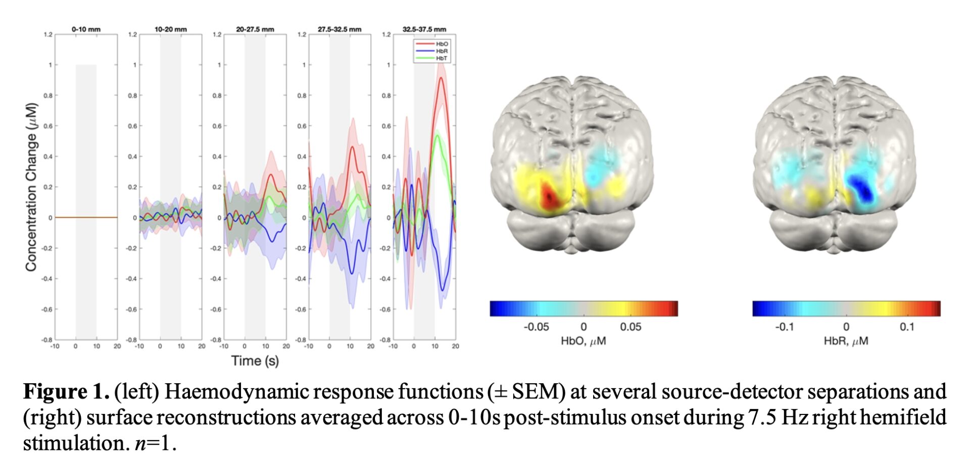
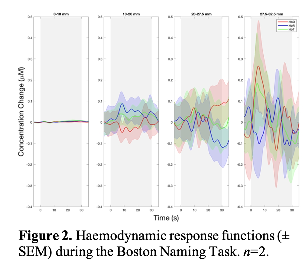
Visual stimulation elicited a clear, delayed rise in oxygenated haemoglobin (HbO; Figure 1; peak ~13.2s from stimulus onset) with a concurrent decrease in deoxygenated haemoglobin (HbR) in the contralateral hemisphere to the stimulated hemifield. The BNT yielded an immediate rise in HbO (Figure 2) only evident in longer source-detector separations, more likely to sample the brain.
Conclusion
A robust response to visual stimulation was observed, whereas the BNT may be insufficiently demanding. These results show promise that NIRS can detect differences between people with dementia and controls. Future work in these populations will test this hypothesis.