
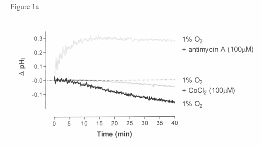
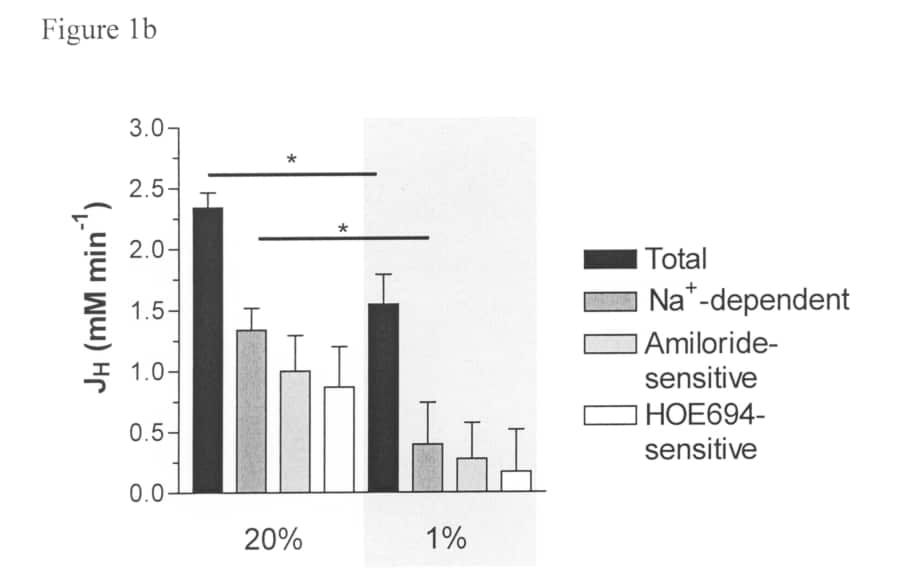
As articular cartilage is avascular, their chondrocytes experience low O2 levels. Values can fall further in chronic disease states. Hypoxia perturbs cell function, inhibiting metabolism and matrix synthesis. It also perturbs ion homeostasis (Gibson et al. 2008). Chondrocyte intracellular pH (pHi) is a powerful regulator of matrix synthesis. Acid extrusion, largely mediated by Na+-H+ exchange (NHE) (Wilkins et al. 2000), is inhibited when cells are incubated for several hours at low O2, leading to intracellular acidification (Milner et al 2006). O2-dependent changes in levels of reactive oxygen species (ROS) appear to underlie this effect. The rapidity with which hypoxia exerts these effects is important both mechanistically and for the consequence of short term hypoxia on joint integrity, but it has not been characterised. Cartilage slices were taken from bovine and equine fetlock joints of animals humanely killed for other purposes. Chondrocytes, isolated by collagenase digestion, were incubated at 20% or 1% O2 at 37oC. pHi was determined fluorimetrically with BCECF (Wilkins & Hall, 1992), using NH4Cl to alter pHi. ROS levels were determined using DCFH. Salines were buffered with HEPES (10 mM), in nominal absence of CO2 / HCO3– and equilibrated at the required O2 tension by incubation in tonometers. Steady state pHi was maintained for periods of up to 40 min (ΔpHi = 0.10±0.04, n=3). A step change to 1% O2 caused a decline in pHi within 10min, and the fall continued throughout the recording period (Figure 1a). At 1% O2 in the presence of CoCl2 (100μM), fall in pHi was largely abolished; at 1% O2 with the mitochondrial complex III inhibitor antimycin A (100µM), a rapid alkalinisation of pHi was observed. ROS levels decreased in response to hypoxia (to 60±15% control values, means+SEM, n=3), but were maintained by treatment with either Co2+ or antimycin A. Recovery from ammonium-imposed intracellular acidification was also slowed within 10min under hypoxia, via reduction in the Na+-dependent / HOE694-sensitive component of the acid flux, consistent with a reduction in NHE activity (Figure 1b). These findings show that hypoxia has rapid effects on chondrocyte function, acidifying cells via NHE inhibition, correlating with alteration in ROS levels. Aside from their obvious implications for chondrocyte homeostasis and cartilage integrity, the rapidity of the response observed here may be of relevance in the context of other hypoxic tissues such as the ischemic myocardium.
University of Cambridge (2008) Proc Physiol Soc 11, PC163
Poster Communications: Rapid effects of hypoxia on pH homeostasis in articular chondrocytes
J. S. Gibson1, T. P. Fairfax2, R. J. Wilkins2, P. I. Milner3
1. Veterinary Medicine, University of Cambridge, Cambridge, United Kingdom. 2. Physiology, Anatomy and Genetics, University of Oxford, Oxford, Ox, United Kingdom. 3. Faculty of Veterinary Science, University of Liverpool, Liverpool, United Kingdom.
View other abstracts by:
Where applicable, experiments conform with Society ethical requirements.