
Insulin-like growth factor-1 (IGF-1) plays important roles in numerous physiological processes. IGF-1 and its receptor (IGF-1R) are present in rat heart, consistent with IGF-1 regulating growth and hypertrophy in the developing heart in an autocrine or paracrine manner. IGF-1 induces cardiac hypertrophy in vitro and in vivo; protective and anti-apoptotic properties for this growth factor have also been described. Changes in intracellular Ca2+ concentration, [Ca2+]i after IGF-1 estimulation were visualized in single cardiomyocytes preloaded with Fluo3-AM. Increases of relative fluorescence represent an increase of free cytosolic [Ca2+]. In cultured rat cardiomyocytes, IGF-1 induced a fast and transient increase in Ca2+i levels apparent both in the nucleus and cytosol, releasing this ion from intracellular stores through an inositol 1,4,5-trisphosphate (IP3 )-dependent signaling pathway. Intracellular IP3 levels increased after IGF-1 stimulation in both the presence and absence of extracellular Ca2+. The basal mass of IP3 was~20 pmol/mg of protein; IGF-1 stimulation significantly increased this value to 90 and 130 pmol/mg of protein (4.5- and 6.5-fold increase) for cardiomyocytes incubated in Ca2+-free and Ca2+-containing resting media, respectively. Data were analyzed by analysis of variance and comparisons between groups were performed using a protected Tukey’s t test. A value of p<0.05 was set as the limit of statistical significance. A different spatial distribution of both PLC isoforms and IP3 receptor isoforms in cardiomyocytes was found. Ryanodine did not prevent the IGF-1-induced increase of Ca2+i levels but inhibited the basal and spontaneous Ca2+i oscillations observed when cardiac myocytes were incubated in Ca2+-containing resting media. Spatial analysis of fluorescence images of IGF-1-stimulated cardiomyocytes incubated in Ca2+-containing resting media showed an early increase in Ca2+i, initially localized in the nucleus. ROI analysis clearly showed that extracellular Ca2+ slowed down the cytosolic but not the nuclear increase in [Ca2+] stimulated by IGF-1. Rates of rise of the nuclear Ca2+ transients in the presence and absence of external Ca2+ were 32.3 ± 7.6 and 38.6 ± 8.1 (Δf/f per s) (n=4), respectively. However, rates of rise of Ca2+ transients in the cytosol were 3.9 ± 1.4 and 20.1 ± 5.3 (Δf/f per s) (n =10), respectively. Calcium imaging suggested that part of the Ca2+ released by stimulation with IGF-1 was initially contained in the perinuclear region. The IGF-1-induced increase on Ca2+i levels was prevented by 1,2-bis(2-aminophenoxy)ethane-N,N,N,N-tetraacetic acid-AM, 100 μM (n= 5), thapsigargin 1 μM (n=4), xestospongins B 10 μM (n=3) and C 100 μM (n=7), 2-aminoethoxy diphenyl borate 20 μM (n=3), U-73122 50 μM (n=8), pertussis toxin 1 μg/ml (n= 7), and βARKct (an adenovirus-transduced peptide inhibitor of Gβγsignalling) (MOI 300) (n=7). Pertussis toxin and βARKct also prevented the IGF-1-dependent IP3 mass increase. Genistein (100 μM)treatment largely decreased the IGF-1- induced changes in both Ca2+i and IP3. LY29402 (but not PD98059) also prevented the IGF-1-dependent Ca2+ increase. βARKct, pertussis toxin and U73122 prevented the IGF-1-dependent induction of ERKs but not protein kinase B (PKB). We conclude that IGF-1 increases Ca2+ levels in cultured cardiac myocytes through a G βγ subunit of a pertussis toxin-sensitive G protein-PI3K-phospholipase C signaling pathway that involves participation of IP3 and perinuclear intracellular calcium store. Finally, this new signaling pathways is necessary for IGF-1-dependent ERKs activation in cardiac myocyte.
King's College London (2005) J Physiol 565P, C76
Communications: Rapid effects of Insulin-like Growth Factor-1 on Nuclear and Cytosolic Calcium in Cultured Rat Cardiac Myocytes
Ibarra Iribarren, Cristian Andres; Estrada, Manuel ; Carrasco, Loreto ; Chiong, Mario ; Jaimovich, Enrique ; Lavandero, Sergio ;
1. FONDAP Center for Molecular Studies of the Cell, University of Chile, Santiago, Chile. 2. Laboratory of Molecular Hermeneutics, Yale School of Medicine, New Haven, NY, USA.
View other abstracts by:
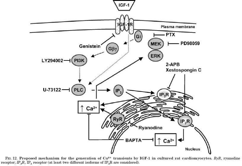
Figure. Proposed mechanism for the generation of Ca2_ transients by IGF-1 in cultured rat cardiomyocytes. RyR ryanodine receptor; IP3R IP3 receptor (at least two different isoforms of IP3R are considered).
Where applicable, experiments conform with Society ethical requirements.