
Myocardial fibre and sheet architecture both influence the dynamics of propagation of the action potential (1,2), and, in addition, both are important determinants in the mechanism of myocardial thickening during contraction (3, 4). Diffusion Tensor – Magnetic Resonance Imaging (DT-MRI) has been validated as a method to extract fibre and sheet architecture (4). DT-MRI studies have shown that cardiac sheet structure exists as two populations, angled at -55° and +35° to the cardiac short-axis plane, and these sheet populations are variable in distribution between canine individuals (4). The abutments of the two sheet populations are elongated circumferentially, around the left-ventricle, and are regions of large spatial gradients in sheet angle. We examined sheet abutments in blocks of left-ventricle from DT-MRI datasets. Five cuboid regions (~30 x 20 x 5 voxels, of dimensions 0.3125 x 0.3125 x 0.9 mm) were selected from three canine hearts. Regions of rapid sheet angle change with distance were selected from throughout the left ventricle. The fibre helix, fibre transverse and sheet angles were extracted and visualised for these regions, and the fibre-orientation visualised in three dimensions. Visualisations of fibre-orientation across positive to negative sheet transitions did not reveal increased fibre-angle divergence across these zones (Figure 1). Quantitative analysis of the fibre angle divergence across sheet angle transitions was carried out on selected rows of voxels from the myocardial blocks. No consistent significant differences in mean voxel-to-voxel divergence of fibre helix angle, fibre transverse angle, primary eigenvalue or vector cosines were found across the sheet angle transition (at p = 0.01). We conclude that the regular helical conformation of cardiac myofibre orientation is maintained across positive and negative sheet abutments. This has implications for the nature of fibre merging between positive and negative sheet populations (4) which must occur in a highly organised and regular fashion. This is important given the variable location of the positive and negative sheet population between individuals.
Life Sciences 2007 (2007) Proc Life Sciences, PC359
Poster Communications: Regular change in canine myocardial fibre orientation across transitions between positive and negative orientated cardiac sheets
S. H. Gilbert1, J. Y. Hodgson1, A. V. Holden1
1. Institute of Membrane and Systems Biology and Cardiovascular Research Institute, University of Leeds, Leeds, West Yorkshire, United Kingdom.
View other abstracts by:
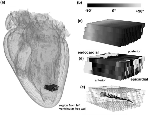
(a) Cardiac location of selected block. (b) Smooth change in the fibre helix angle during (c) discontinuous change in the sheet angle (positive to negative sheet transition) and (d) combined smooth changes in fibre orientation (black rods) with discontinuous changes in sheet angle (shaded grid). (e) Monochrome angular scale.
Where applicable, experiments conform with Society ethical requirements.