
Background: The carotid body (CB) detects and translates blood-borne stimuli into patterns of neural discharge to initiate corrective chemoreflexes. Elevated CB activity has been implicated in the pathophysiology of cardiorespiratory diseases including hypertension and cardiac arrhythmia (1). Hypoxia increases CB activity and the steady-state magnitude of its sensory output in an intensity-dependent manner. However, little is known about the patterning of CB sensory discharge. It is hypothesised that a shift from a random action potential pattern to a more regular pattern determines the physiological and pathological responses elicited by different intensities of hypoxia.
Aims: This study sought to analyse differences in the randomness and variability of action potential (AP) firing patterns recorded from CB chemoafferent fibres during graded hypoxic stimulation.
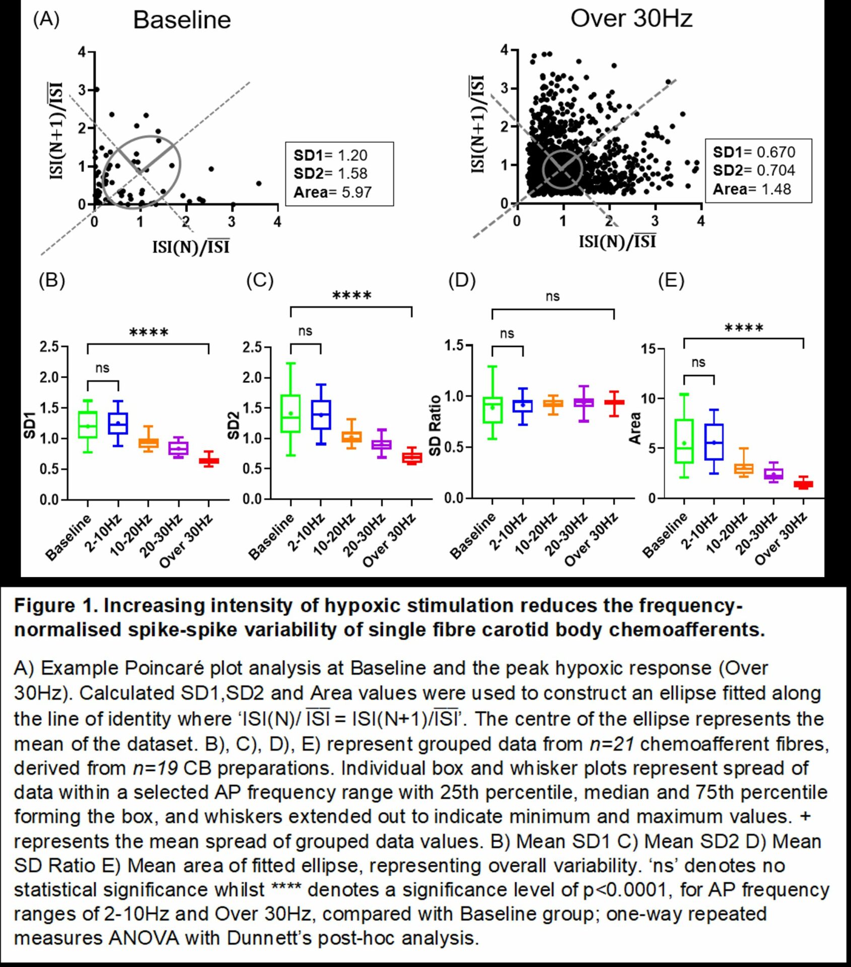
Methods: Ex vivo extracellular nerve recordings were made from Wistar rat CBs (male, aged 5-20 weeks, n=19), removed under terminal anaesthesia (2.5-4% isoflurane in O2 at 1.5L.min-1). Single-fibre activity (n=21) was distinguished using Spike2 (version 9.05) software. Data was grouped into five hypoxic intensities defined by their mean AP frequency range; 0-2Hz (baseline), 2-10Hz, 10-20Hz, 20-30Hz, over 30Hz. Instantaneous frequency (IF) and Poincaré plot analysis, which characterised inter-spike interval (ISI) distribution, were used as quantitative measures of AP patterning. Values are expressed as mean ± S.E.M, compared using one-way ANOVA followed by Dunnett’s post-hoc analysis.
Results: Compared to baseline, the IF distribution of chemoafferent firing during severe hypoxia (over 30Hz) was not random (IF SD:Mean ratio; 2.4±0.127 vs 0.535±0.034, p<0.001). Standard measures of dispersion obtained from frequency-normalised Poincaré plots, SD1 and SD2, were both significantly reduced in severe hypoxia (Figure 1 A-D) leading to a lower overall variability between successive ISIs (Area of ellipse; baseline: 5.52±0.532AU vs over 30Hz:1.401±0.092AU, p<0.001) (Figure 1A & E). No changes between baseline and mild hypoxia (2-10Hz) were observed in the randomness or variability in AP firing, indicating that a minimum intensity is required to modify CB sensory patterning. 2-10Hz, but not over 30Hz hypoxia significantly increased the maximum IF compared to baseline (baseline:109.9±14.4Hz vs 2-10Hz:160.0±8.55Hz, p<0.01), thus suggesting that some features of CB sensory discharge are not correlated with stimulus intensity.
Conclusion: This data shows that higher intensities of hypoxia modulate CB sensory patterning, most notably by becoming less random and more regular. Investigation of their functional significance and the impact of disease states may establish CB discharge patterning as an alternative therapeutic target. Future studies may also evaluate CB chemoafferent responses to other stimuli (hypercapnia, acidosis, mitochondrial inhibition) which may generate different chemoafferent patterns compared to hypoxia.
Sensory Signals (The Royal College of Physicians, London, UK) (2022) Proc Physiol Soc 50, C20
Poster Communications: Severe hypoxia reduces the randomness and variability of carotid body (CB) sensory patterning
Natasha Williams1, Andrew Holmes1, 2, 3, Prem Kumar1, 2, 3, Clare Ray1, 3, 4, Andrew Coney1, 3, 5
1University of Birmingham, Birmingham, United Kingdom 2Institute of Clinical Sciences, Birmingham, United Kingdom 3Birmingham Arterial Chemoreceptor and Hypoxia Group, Birmingham, United Kingdom 4School of Biomedical Sciences, Birmingham, United Kingdom 5Institute of Cardiovascular Sciences, University of Birmingham, United Kingdom
View other abstracts by:
Where applicable, experiments conform with Society ethical requirements.